Cryophotolysis of Caged Compounds: A Technique for Trapping Intermediate States in Protein Crystals
Ursby, T., Weik, M., Fioravanti, E., Delarue, M., Goeldner, M., Bourgeois, D.(2002) Acta Crystallogr D Biol Crystallogr 58: 607
- PubMed: 11914484 Search on PubMed
- DOI: https://doi.org/10.1107/s0907444902002135
- Primary Citation Related Structures:
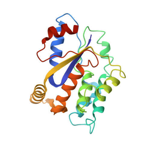
1GSI, 1GTV - PubMed Abstract:
Caged compounds in combination with protein crystallography represent a valuable tool in studies of enzyme reaction intermediates. To date, photochemical triggering of reactions has been performed close to room temperature. Synchronous reaction initiation has only been achieved with enzymes of relatively slow turnover (<0.1 s(-1)) and caged compounds of high quantum yield. Here X-ray crystallography and microspectrophotometry were used to provide evidence that (nitrophenyl)ethyl (NPE) ester bonds can be photolyzed by UV light at cryotemperatures. NPE-caged ATP in flash-cooled crystals of Mycobacterium tuberculosis thymidylate kinase was photolyzed successfully at 100-150 K as assessed by the structural observation of ATP-dependent enzymatic conversion of TMP to TDP after temporarily warming the crystals to room temperature. A new method is proposed in which cryo-photolysis combined with temperature-controlled protein crystallography can be used to trap reaction intermediates even in some of the fastest enzymes and/or when only compounds of low quantum yield are available. Raising the temperature after cryophotolysis may allow a transition barrier to be passed and an intermediate to accumulate in the crystal. A comparable method has only been used so far with proteins displaying endogenous photosensitivity. The approach described here opens the way to studying the reaction mechanisms of a much larger number of crystalline enzymes. Furthermore, it is shown that X-ray-induced radiolysis of caged compounds occurs if high-intensity synchrotron beamlines are used. This caveat should be taken into account when deriving data-collection protocols. It could also be used potentially as a way to trigger reactions.
- LCCP, UMR 9015, Institut de Biologie Structurale, 41 Avenue Jules Horowitz, 38027 Grenoble CEDEX 1, France.
Organizational Affiliation: