1GTT | pdb_00001gtt

CRYSTAL STRUCTURE OF HPCE


Experimental Data Snapshot

  • Method: X-RAY DIFFRACTION
  • Resolution: 1.70 Å
  • R-Value Free: 
    0.252 (Depositor), 0.266 (DCC) 
  • R-Value Work: 
    0.216 (Depositor) 
  • R-Value Observed: 
    0.217 (Depositor) 

wwPDB Validation 3D Report Full Report

Validation slider image for 1GTT

This is version 1.4 of the entry. See complete history

Literature

The Crystal Structure of Hpce, a Bifunctional Decarboxylase/Isomerase with a Multifunctional Fold.

Tame, J.R.H.Namba, K.Dodson, E.J.Roper, D.I.

(2002) Biochemistry 41: 2982

  • DOI: https://doi.org/10.1021/bi015717t
  • Primary Citation Related Structures: 
    1GTT

  • PubMed Abstract: 

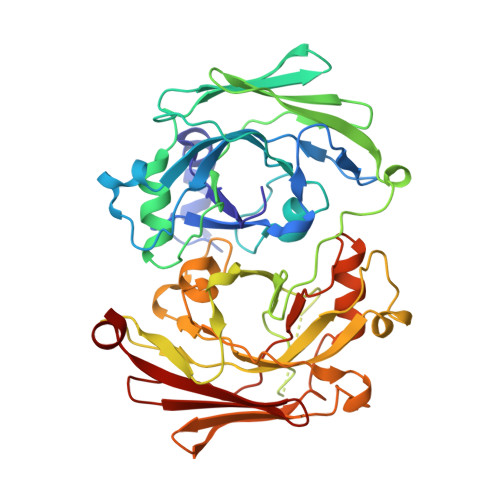
    The structure of the bifunctional enzyme HpcE (OPET decarboxylase/HHDD isomerase) from Escherichia coli shows that the protein consists of highly similar N and C terminal halves. Sequence matches suggest that this fold is widespread among different species, including man. Many of these homologues are uncharacterized but apparently connected with the metabolism of aromatic compounds. The domain shows similar topology to the C terminal domain of fumarylacetoacetate hydrolase (FAH), a functionally related enzyme, despite lacking significant overall sequence similarity. HpcE is known to catalyze two rather different reactions, and comparisons with FAH allow some tentative conclusions to be drawn about the active sites. Key mutations within the active site apparently allow enzymes with this fold to carry out a variety chemical processes.


  • Organizational Affiliation
    • Protein Design Laboratory, Yokohama City University, Suehiro 1-7-29, Kanagawa 230-0045, Japan. jtame@tsurumi.yokohama-cu.ac.jp

Macromolecule Content 

  • Total Structure Weight: 188.78 kDa 
  • Atom Count: 14,274 
  • Modeled Residue Count: 1,684 
  • Deposited Residue Count: 1,716 
  • Unique protein chains: 1

Macromolecules

Find similar proteins by:|  3D Structure
Entity ID: 1
MoleculeChains  Sequence LengthOrganismDetailsImage
4-HYDROXYPHENYLACETATE DEGRADATION BIFUNCTIONAL ISOMERASE/DECARBOXYLASE
A, B, C, D
429Escherichia coliMutation(s): 0 
EC: 5.3.3.10 (PDB Primary Data), 4.1.1 (PDB Primary Data), 4.1.1.68 (UniProt)
UniProt
Find proteins for P37352 (Escherichia coli)
Explore P37352 
Go to UniProtKB:  P37352
Entity Groups
Sequence Clusters30% Identity50% Identity70% Identity90% Identity95% Identity100% Identity
UniProt GroupP37352
Sequence Annotations
Expand
Reference Sequence

Experimental Data & Validation

Experimental Data

  • Method: X-RAY DIFFRACTION
  • Resolution: 1.70 Å
  • R-Value Free:  0.252 (Depositor), 0.266 (DCC) 
  • R-Value Work:  0.216 (Depositor) 
  • R-Value Observed: 0.217 (Depositor) 
Space Group: P 21 21 21
Unit Cell:
Length ( Å )Angle ( ˚ )
a = 126.084α = 90
b = 138.201β = 90
c = 103.973γ = 90
Software Package:
Software NamePurpose
REFMACrefinement
MARdata reduction
DENZOdata reduction
SCALEPACKdata scaling
MLPHAREphasing

Structure Validation

View Full Validation Report


Entry History 

Deposition Data

Revision History  (Full details and data files)

  • Version 1.0: 2002-03-08
    Type: Initial release
  • Version 1.1: 2011-05-08
    Changes: Version format compliance
  • Version 1.2: 2011-07-13
    Changes: Version format compliance
  • Version 1.3: 2019-07-24
    Changes: Advisory, Data collection
  • Version 1.4: 2024-11-13
    Changes: Advisory, Data collection, Database references, Derived calculations, Other, Structure summary