Biochemical and Crystallographic Studies of the met144Ala, Asp92Asn and His254Phe Mutants of the Nitrite Reductase from Alcaligenes Xylosoxidans Provide Insight Into the Enzyme Mechanism.
Ellis, M.J., Prudencio, M., Dodd, F.E., Strange, R.W., Sawers, G., Eady, R.R., Hasnain, S.S.(2002) J Mol Biology 316: 51
- PubMed: 11829502 Search on PubMed
- DOI: https://doi.org/10.1006/jmbi.2001.5304
- Primary Citation Related Structures:
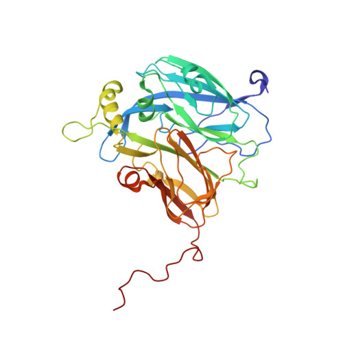
1GS6, 1GS7, 1GS8 - PubMed Abstract:
Dissimilatory nitrite reductase catalyses the reduction of nitrite (NO(2)(-)) to nitric oxide (NO). Copper-containing nitrite reductases contain both type 1 and type 2 Cu sites. Electron transfer from redox partners is presumed to be mediated via the type 1 Cu site and used at the catalytic type 2 Cu centre along with the substrate nitrite. At the type 2 Cu site, Asp92 has been identified as a key residue in substrate utilisation, since it hydrogen bonds to the water molecule at the nitrite binding site. We have also suggested that protons enter the catalytic site via Asp92, through a water network that is mediated by His254. The role of these residues has been investigated in the blue copper nitrite reductase from Alcaligenes xylosoxidans (NCIMB 11015) by a combination of point mutation, enzymatic activity measurement and structure determination.In addition, it has been suggested that the enzyme operates via an ordered mechanism where an electron is transferred to the type 2 Cu site largely when the second substrate nitrite is bound and that this is controlled via the lowering of the redox potential of the type 2 site when it is loaded with nitrite. Thus, a small perturbation of the type 1 Cu site should result in a significant effect on the activity of the enzyme. For this reason a mutation of Met144, which is the weakest ligand of the type 1 Cu, is investigated. The structures of H254F, D92N and M144A have been determined to 1.85 A, 1.9 A and 2.2 A resolution, respectively. The D92N and H254F mutants have negligible or no activity, while the M144A mutant has 30 % activity of the native enzyme. Structural and spectroscopic data show that the loss of activity in H254F is due to the catalytic site being occupied by Zn while the loss/reduction of activity in D92N/M144A are due to structural reasons. The D92N mutation results in the loss of the Asp92 hydrogen bond to the Cu-ligated water. Therefore, the ligand is no longer able to perform proton abstraction. Even though the loss of activity in H254F is due to lack of catalytic Cu, the mutation does cause the disruption of the water network, confirming its key role in proton channel. The structure of the H254F mutant is the first case where full occupancy Zn at the type 2 Cu site is observed, but despite the previously noted similarity of this site to the carbonic anhydrase catalytic site, no carbonic anhydrase activity is observed. The H254F and D92N mutant structures provide, for the first time, observation of surface Zn sites which may act as a Zn sink and prevent binding of Zn at the catalytic Cu site in the native enzyme.
- Faculty of Applied Science, De Montfort University, Leicester, LE1 9BH, UK.
Organizational Affiliation: