1GRH | pdb_00001grh

INHIBITION OF HUMAN GLUTATHIONE REDUCTASE BY THE NITROSOUREA DRUGS 1,3-BIS(2-CHLOROETHYL)-1-NITROSOUREA AND 1-(2-CHLOROETHYL)-3-(2-HYDROXYETHYL)-1-NITROSOUREA


Experimental Data Snapshot

  • Method: X-RAY DIFFRACTION
  • Resolution: 3.00 Å

wwPDB Validation 3D Report Full Report

Validation slider image for 1GRH

This is version 1.4 of the entry. See complete history

Literature

Inhibition of human glutathione reductase by the nitrosourea drugs 1,3-bis(2-chloroethyl)-1-nitrosourea and 1-(2-chloroethyl)-3-(2-hydroxyethyl)-1-nitrosourea. A crystallographic analysis.

Karplus, P.A.Krauth-Siegel, R.L.Schirmer, R.H.Schulz, G.E.

(1988) Eur J Biochem 171: 193-198

  • DOI: https://doi.org/10.1111/j.1432-1033.1988.tb13775.x
  • Primary Citation Related Structures: 
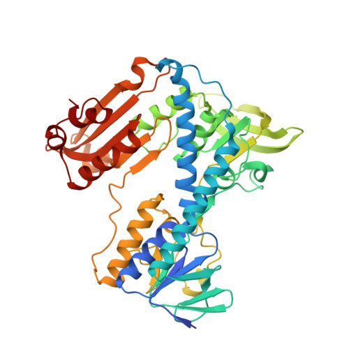
    1GRH

  • PubMed Abstract: 

    Glutathione reductase from human erythrocytes was inhibited by incubation with the drugs 1,3-bis(2-chloroethyl)-1-nitrosourea (BCNU) and 1-(2-chloroethyl)-3-(2-hydroxyethyl)-1-nitrosourea (HeCNU) under quasi-physiological conditions. For reference purposes, iodoacetamide was used for inactivating alkylation of the enzyme. In each case the modified glutathione reductase was crystallized and its structure determined. These analyses showed that in all experiments the enzyme had reacted at the distal sulfur, that is at the thiol of Cys-58, and virtually nowhere else in the visible structure. The electron density of the HeCNU derivative at 0.3 nm resolution is consistent with a 2-hydroxyethyl group. This alkyl moiety has recently been identified by chemical analysis [Schirmer, R. H., Schöllhammer, T., Eisenbrand, G. and Krauth-Siegel, R. L. (1987) Free Radical Res. Commun. 3, 3-12]. The 0.2 nm resolution electron-density map of the BCNU-derivatized enzyme cannot be explained by a 2-hydroxyethyl group. Instead the modification appears as a carbamoyl moiety containing at least five non-hydrogen atoms. In this derivative the distal cysteine is forced into an unusual conformation.


  • Organizational Affiliation
    • Institut für Organische Chemie und Biochemie, Universität Freiburg i. Br., Federal Republic of Germany.

Macromolecule Content 

  • Total Structure Weight: 52.56 kDa 
  • Atom Count: 4,080 
  • Modeled Residue Count: 461 
  • Deposited Residue Count: 478 
  • Unique protein chains: 1

Macromolecules

Find similar proteins by:|  3D Structure
Entity ID: 1
MoleculeChains  Sequence LengthOrganismDetailsImage
GLUTATHIONE REDUCTASE478Homo sapiensMutation(s): 0 
EC: 1.6.4.2 (PDB Primary Data), 1.8.1.7 (UniProt)
UniProt & NIH Common Fund Data Resources
Find proteins for P00390 (Homo sapiens)
Explore P00390 
Go to UniProtKB:  P00390
PHAROS:  P00390
GTEx:  ENSG00000104687 
Entity Groups
Sequence Clusters30% Identity50% Identity70% Identity90% Identity95% Identity100% Identity
UniProt GroupP00390
Sequence Annotations
Expand
Reference Sequence

Small Molecules

Ligands 3 Unique
IDChains Name / Formula / InChI Key2D Diagram3D Interactions
FAD

Query on FAD


Download:Ideal Coordinates CCD File
C [auth A]FLAVIN-ADENINE DINUCLEOTIDE
C27 H33 N9 O15 P2
VWWQXMAJTJZDQX-UYBVJOGSSA-N
PO4

Query on PO4


Download:Ideal Coordinates CCD File
B [auth A]PHOSPHATE ION
O4 P
NBIIXXVUZAFLBC-UHFFFAOYSA-K
EOH

Query on EOH


Download:Ideal Coordinates CCD File
D [auth A]ETHANOL
C2 H6 O
LFQSCWFLJHTTHZ-UHFFFAOYSA-N

Experimental Data & Validation

Experimental Data

  • Method: X-RAY DIFFRACTION
  • Resolution: 3.00 Å
Space Group: B 1 1 2
Unit Cell:
Length ( Å )Angle ( ˚ )
a = 119.8α = 90
b = 84.5β = 90
c = 63.2γ = 58.7

Structure Validation

View Full Validation Report


Entry History 

Deposition Data

Revision History  (Full details and data files)

  • Version 1.0: 1994-01-31
    Type: Initial release
  • Version 1.1: 2011-06-03
    Changes: Version format compliance
  • Version 1.2: 2011-07-13
    Changes: Version format compliance
  • Version 1.3: 2011-07-27
    Changes: Other
  • Version 1.4: 2024-06-05
    Changes: Data collection, Database references, Derived calculations, Other