1GR2

STRUCTURE OF A GLUTAMATE RECEPTOR LIGAND BINDING CORE (GLUR2) COMPLEXED WITH KAINATE


Experimental Data Snapshot

  • Method: X-RAY DIFFRACTION
  • Resolution: 1.90 Å
  • R-Value Free: 0.273 
  • R-Value Work: 0.216 
  • R-Value Observed: 0.216 

wwPDB Validation   3D Report Full Report


This is version 1.3 of the entry. See complete history


Literature

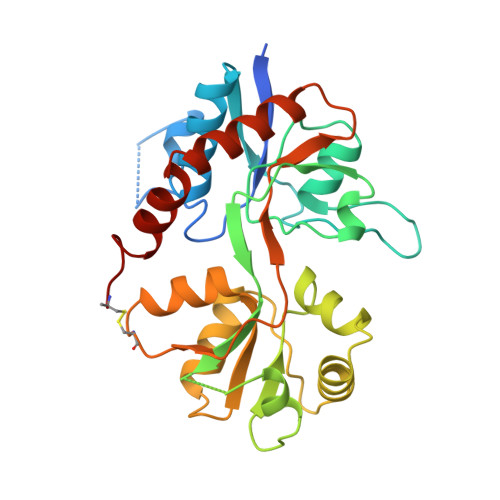
Structure of a glutamate-receptor ligand-binding core in complex with kainate.

Armstrong, N.Sun, Y.Chen, G.Q.Gouaux, E.

(1998) Nature 395: 913-917

  • DOI: https://doi.org/10.1038/27692
  • Primary Citation of Related Structures:  
    1GR2

  • PubMed Abstract: 

    Ionotropic glutamate receptors (iGluRs) mediate excitatory synaptic transmission in vertebrates and invertebrates through ligand-induced opening of transmembrane ion channels. iGluRs are segregated into three subtypes according to their sensitivity to the agonists AMPA (alpha-amino-3-hydroxy-5-methyl-4-isoxazole propionic acid), kainate (a structural analogue of glutamate) or NMDA (N-methyl-D-aspartate). iGluRs are important in the development and function of the nervous system, are essential in memory and learning, and are either implicated in or have causal roles in dysfunctions ranging from Alzheimer's, Parkinson's and Huntington's diseases, schizophrenia, epilepsy and Rasmussen's encephalitis to stroke. Development of iGluR agonists and antagonists has been hampered by a lack of high-resolution structural information. Here we describe the crystal structure of an iGluR ligand-binding region in a complex with the neurotoxin (agonist) kainate. The bilobed structure shows the determinants of receptor-agonist interactions and how ligand-binding specificity and affinity are altered by remote residues and the redox state of the conserved disulphide bond. The structure indicates mechanisms for allosteric effector action and for ligand-induced channel gating. The information provided by this structure will be essential in designing new ligands.


  • Organizational Affiliation

    Department of Biochemistry and Molecular Biophysics, Columbia University, New York, New York 10032, USA.


Macromolecules
Find similar proteins by:  (by identity cutoff)  |  3D Structure
Entity ID: 1
MoleculeChains Sequence LengthOrganismDetailsImage
PROTEIN (GLUTAMATE RECEPTOR 2)279Rattus norvegicusMutation(s): 0 
UniProt
Find proteins for P19491 (Rattus norvegicus)
Explore P19491 
Go to UniProtKB:  P19491
Entity Groups  
Sequence Clusters30% Identity50% Identity70% Identity90% Identity95% Identity100% Identity
UniProt GroupP19491
Sequence Annotations
Expand
  • Reference Sequence
Small Molecules
Ligands 1 Unique
IDChains Name / Formula / InChI Key2D Diagram3D Interactions
KAI
Query on KAI

Download Ideal Coordinates CCD File 
B [auth A]3-(CARBOXYMETHYL)-4-ISOPROPENYLPROLINE
C10 H15 N O4
VLSMHEGGTFMBBZ-OOZYFLPDSA-N
Binding Affinity Annotations 
IDSourceBinding Affinity
KAI BindingDB:  1GR2 Ki: min: 1.8, max: 1.22e+4 (nM) from 11 assay(s)
EC50: min: 1.06e+4, max: 3.80e+5 (nM) from 8 assay(s)
Experimental Data & Validation

Experimental Data

  • Method: X-RAY DIFFRACTION
  • Resolution: 1.90 Å
  • R-Value Free: 0.273 
  • R-Value Work: 0.216 
  • R-Value Observed: 0.216 
  • Space Group: P 1 21 1
Unit Cell:
Length ( Å )Angle ( ˚ )
a = 43.22α = 90
b = 63.07β = 92.99
c = 46.32γ = 90
Software Package:
Software NamePurpose
DENZOdata reduction
SCALEPACKdata scaling
SOLVEphasing
X-PLORrefinement

Structure Validation

View Full Validation Report


Entry History 

Deposition Data

Revision History  (Full details and data files)

  • Version 1.0: 1998-12-09
    Type: Initial release
  • Version 1.1: 2008-04-01
    Changes: Version format compliance
  • Version 1.2: 2011-07-13
    Changes: Version format compliance
  • Version 1.3: 2017-08-16
    Changes: Refinement description, Source and taxonomy