1GQF

Crystal structure of human procaspase-7


Experimental Data Snapshot

  • Method: X-RAY DIFFRACTION
  • Resolution: 2.90 Å
  • R-Value Free: 0.285 
  • R-Value Work: 0.268 
  • R-Value Observed: 0.268 

wwPDB Validation   3D Report Full Report


This is version 1.3 of the entry. See complete history


Literature

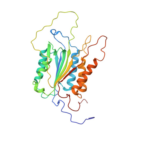
Structural basis for the activation of human procaspase-7.

Riedl, S.J.Fuentes-Prior, P.Renatus, M.Kairies, N.Krapp, S.Huber, R.Salvesen, G.S.Bode, W.

(2001) Proc Natl Acad Sci U S A 98: 14790-14795

  • DOI: https://doi.org/10.1073/pnas.221580098
  • Primary Citation of Related Structures:  
    1GQF

  • PubMed Abstract: 

    Caspases form a family of proteinases required for the initiation and execution phases of apoptosis. Distinct proapoptotic stimuli lead to activation of the initiator caspases-8 and -9, which in turn activate the common executioner caspases-3 and -7 by proteolytic cleavage. Whereas crystal structures of several active caspases have been reported, no three-dimensional structure of an uncleaved caspase zymogen is available so far. We have determined the 2.9-A crystal structure of recombinant human C285A procaspase-7 and have elucidated the activation mechanism of caspases. The overall fold of the homodimeric procaspase-7 resembles that of the active tetrameric caspase-7. Each monomer is organized in two structured subdomains connected by partially flexible linkers, which asymmetrically occupy and block the central cavity, a typical feature of active caspases. This blockage is incompatible with a functional substrate binding site/active site. After proteolytic cleavage within the flexible linkers, the newly formed chain termini leave the cavity and fold outward to form stable structures. These conformational changes are associated with the formation of an intact active-site cleft. Therefore, this mechanism represents a formerly unknown type of proteinase zymogen activation.


  • Organizational Affiliation

    Abteilung Strukturforschung, Max-Planck-Institut für Biochemie, Am Klopferspitz 18a, D-82152 Martinsried, Germany. riedl@biochem.mpg.de


Macromolecules
Find similar proteins by:  (by identity cutoff)  |  3D Structure
Entity ID: 1
MoleculeChains Sequence LengthOrganismDetailsImage
Caspase-7
A, B
265Homo sapiensMutation(s): 1 
Gene Names: CASP7MCH3
EC: 3.4.22.60
UniProt & NIH Common Fund Data Resources
Find proteins for P55210 (Homo sapiens)
Explore P55210 
Go to UniProtKB:  P55210
PHAROS:  P55210
GTEx:  ENSG00000165806 
Entity Groups  
Sequence Clusters30% Identity50% Identity70% Identity90% Identity95% Identity100% Identity
UniProt GroupP55210
Sequence Annotations
Expand
  • Reference Sequence
Experimental Data & Validation

Experimental Data

  • Method: X-RAY DIFFRACTION
  • Resolution: 2.90 Å
  • R-Value Free: 0.285 
  • R-Value Work: 0.268 
  • R-Value Observed: 0.268 
  • Space Group: P 32 2 1
Unit Cell:
Length ( Å )Angle ( ˚ )
a = 90.21α = 90
b = 90.21β = 90
c = 183.02γ = 120
Software Package:
Software NamePurpose
CNSrefinement
MOSFLMdata reduction
SCALAdata scaling
AMoREphasing

Structure Validation

View Full Validation Report


Entry History 

Deposition Data

Revision History  (Full details and data files)

  • Version 1.0: 2002-01-04
    Type: Initial release
  • Version 1.1: 2015-09-16
    Changes: Database references, Derived calculations, Non-polymer description, Other, Refinement description, Structure summary, Version format compliance
  • Version 1.2: 2018-11-07
    Changes: Advisory, Data collection, Database references, Experimental preparation, Source and taxonomy, Structure summary
  • Version 1.3: 2023-12-13
    Changes: Advisory, Data collection, Database references, Derived calculations, Refinement description