1G98 | pdb_00001g98

CRYSTAL STRUCTURE ANALYSIS OF RABBIT PHOSPHOGLUCOSE ISOMERASE COMPLEXED WITH 5-PHOSPHOARABINONATE, A TRANSITION STATE ANALOGUE


Experimental Data Snapshot

  • Method: X-RAY DIFFRACTION
  • Resolution: 1.90 Å
  • R-Value Free: 
    0.236 (Depositor) 
  • R-Value Work: 
    0.211 (Depositor) 

Starting Model: experimental
View more details

wwPDB Validation 3D Report Full Report

Validation slider image for 1G98

This is version 1.4 of the entry. See complete history

Literature

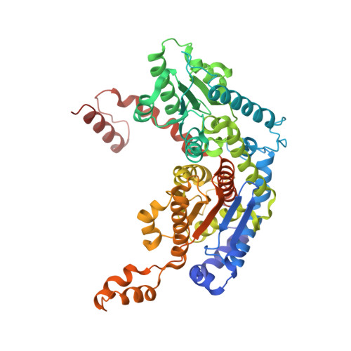
Crystal structure of rabbit phosphoglucose isomerase complexed with 5-phospho-D-arabinonate identifies the role of Glu357 in catalysis.

Jeffery, C.J.Hardre, R.Salmon, L.

(2001) Biochemistry 40: 1560-1566

  • DOI: https://doi.org/10.1021/bi0018483
  • Primary Citation Related Structures: 
    1G98

  • PubMed Abstract: 

    Phosphoglucose isomerase (PGI; E.C. 5.3.1.9) catalyzes the second step in glycolysis, the interconversion of D-glucose-6-phosphate and D-fructose-6-phosphate. We determined the X-ray crystal structure of rabbit PGI complexed with a competitive inhibitor of isomerase activity, 5-phospho-D-arabinonate (5PAA), at 1.9 A resolution. 5PAA is a better mimic of the proposed cis-enediol(ate) intermediate than 6-phospho-D-gluconate, which was used in a previously reported crystal structure of rabbit PGI. The orientation of 5PAA bound in the enzyme active site predicts that active site residue Glu357 is the residue that transfers a proton between C2 and C1 of the proposed cis-enediol(ate) intermediate. Amino acid residues Arg272 and Lys210 are predicted to be involved in stabilizing the negative charge of the intermediate.


  • Organizational Affiliation
    • Laboratory for Molecular Biology, MC567, Department of Biology, University of Illinois at Chicago, Chicago, IL 60607, USA. cjeffery@uic.edu

Macromolecule Content 

  • Total Structure Weight: 126.15 kDa 
  • Atom Count: 9,712 
  • Modeled Residue Count: 1,110 
  • Deposited Residue Count: 1,116 
  • Unique protein chains: 1

Macromolecules

Find similar proteins by:|  3D Structure
Entity ID: 1
MoleculeChains  Sequence LengthOrganismDetailsImage
PHOSPHOGLUCOSE ISOMERASE
A, B
558Oryctolagus cuniculusMutation(s): 0 
EC: 5.3.1.9 (PDB Primary Data), 5.1.3 (UniProt)
UniProt
Find proteins for Q9N1E2 (Oryctolagus cuniculus)
Explore Q9N1E2 
Go to UniProtKB:  Q9N1E2
Entity Groups
Sequence Clusters30% Identity50% Identity70% Identity90% Identity95% Identity100% Identity
UniProt GroupQ9N1E2
Sequence Annotations
Expand
Reference Sequence

Small Molecules

Ligands 1 Unique
IDChains Name / Formula / InChI Key2D Diagram3D Interactions
PA5

Query on PA5


Download:Ideal Coordinates CCD File
C [auth A],
D [auth B]
5-PHOSPHOARABINONIC ACID
C5 H11 O9 P
HNECGPFIYSOYHF-JJYYJPOSSA-N

Experimental Data & Validation

Experimental Data

  • Method: X-RAY DIFFRACTION
  • Resolution: 1.90 Å
  • R-Value Free:  0.236 (Depositor) 
  • R-Value Work:  0.211 (Depositor) 
Space Group: C 2 2 21
Unit Cell:
Length ( Å )Angle ( ˚ )
a = 82.829α = 90
b = 116.554β = 90
c = 271.824γ = 90
Software Package:
Software NamePurpose
CNSrefinement
DENZOdata reduction
SCALEPACKdata scaling
CNSphasing

Structure Validation

View Full Validation Report


Entry History 

Deposition Data

Revision History  (Full details and data files)

  • Version 1.0: 2001-06-01
    Type: Initial release
  • Version 1.1: 2008-04-27
    Changes: Version format compliance
  • Version 1.2: 2011-07-13
    Changes: Version format compliance
  • Version 1.3: 2011-11-16
    Changes: Atomic model
  • Version 1.4: 2023-08-09
    Changes: Data collection, Database references, Derived calculations, Refinement description