1G5H

CRYSTAL STRUCTURE OF THE ACCESSORY SUBUNIT OF MURINE MITOCHONDRIAL POLYMERASE GAMMA


Experimental Data Snapshot

  • Method: X-RAY DIFFRACTION
  • Resolution: 1.95 Å
  • R-Value Free: 0.224 
  • R-Value Work: 0.183 
  • R-Value Observed: 0.183 

wwPDB Validation   3D Report Full Report


This is version 1.3 of the entry. See complete history


Literature

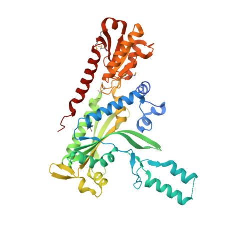
Crystal structure and deletion analysis show that the accessory subunit of mammalian DNA polymerase gamma, Pol gamma B, functions as a homodimer.

Carrodeguas, J.A.Theis, K.Bogenhagen, D.F.Kisker, C.

(2001) Mol Cell 7: 43-54

  • DOI: https://doi.org/10.1016/s1097-2765(01)00153-8
  • Primary Citation of Related Structures:  
    1G5H, 1G5I

  • PubMed Abstract: 

    Polymerase gamma, which replicates and repairs mitochondrial DNA, requires the Pol gamma B subunit for processivity. We determined the crystal structure of mouse Pol gamma B, a core component of the mitochondrial replication machinery. Pol gamma B shows high similarity to glycyl-tRNA synthetase and dimerizes through an unusual intermolecular four-helix bundle. A human Pol gamma B mutant lacking the four-helix bundle failed to dimerize in solution or to stimulate the catalytic subunit Pol gamma A, but retained the ability to bind with Pol gamma A to a primer-template construct, indicating that the functional holoenzyme contains two Pol gamma B molecules. Other mutants retained stimulatory activity but lost the ability to bind folded ssDNA. These results suggest that the Pol gamma B dimer contains distinct sites for Pol gamma A binding, dimerization, and DNA binding.


  • Organizational Affiliation

    Department of Pharmacological Sciences, State University of New York at Stony Brook, Stony Brook, NY 11794, USA.


Macromolecules
Find similar proteins by:  (by identity cutoff)  |  3D Structure
Entity ID: 1
MoleculeChains Sequence LengthOrganismDetailsImage
MITOCHONDRIAL DNA POLYMERASE ACCESSORY SUBUNIT
A, B, C, D
454Mus musculusMutation(s): 6 
Gene Names: DNA POLYMERASE GAMMA SUBUNIT B
UniProt & NIH Common Fund Data Resources
Find proteins for Q9QZM2 (Mus musculus)
Explore Q9QZM2 
Go to UniProtKB:  Q9QZM2
IMPC:  MGI:1354947
Entity Groups  
Sequence Clusters30% Identity50% Identity70% Identity90% Identity95% Identity100% Identity
UniProt GroupQ9QZM2
Sequence Annotations
Expand
  • Reference Sequence
Experimental Data & Validation

Experimental Data

  • Method: X-RAY DIFFRACTION
  • Resolution: 1.95 Å
  • R-Value Free: 0.224 
  • R-Value Work: 0.183 
  • R-Value Observed: 0.183 
  • Space Group: P 21 21 21
Unit Cell:
Length ( Å )Angle ( ˚ )
a = 96.623α = 90
b = 133.422β = 90
c = 135.038γ = 90
Software Package:
Software NamePurpose
SHAKEmodel building
SnBphasing
SOLVEphasing
SHARPphasing
REFMACrefinement
MARMADdata reduction
DENZOdata reduction
SCALEPACKdata scaling

Structure Validation

View Full Validation Report


Entry History 

Deposition Data

Revision History  (Full details and data files)

  • Version 1.0: 2001-03-14
    Type: Initial release
  • Version 1.1: 2008-04-27
    Changes: Version format compliance
  • Version 1.2: 2011-07-13
    Changes: Non-polymer description, Version format compliance
  • Version 1.3: 2017-10-04
    Changes: Refinement description