1G1E | pdb_00001g1e

NMR STRUCTURE OF THE HUMAN MAD1 TRANSREPRESSION DOMAIN SID IN COMPLEX WITH MAMMALIAN SIN3A PAH2 DOMAIN


Experimental Data Snapshot

  • Method: SOLUTION NMR
  • Conformers Calculated: 25 
  • Conformers Submitted: 15 
  • Selection Criteria: structures with favorable non-bond energy,structures with the least restraint violations 

wwPDB Validation 3D Report Full Report

Validation slider image for 1G1E

This is version 1.4 of the entry. See complete history

Literature

Solution structure of the interacting domains of the Mad-Sin3 complex: implications for recruitment of a chromatin-modifying complex.

Brubaker, K.Cowley, S.M.Huang, K.Loo, L.Yochum, G.S.Ayer, D.E.Eisenman, R.N.Radhakrishnan, I.

(2000) Cell 103: 655-665

  • DOI: https://doi.org/10.1016/s0092-8674(00)00168-9
  • Primary Citation Related Structures: 
    1G1E

  • PubMed Abstract: 

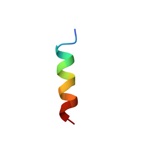
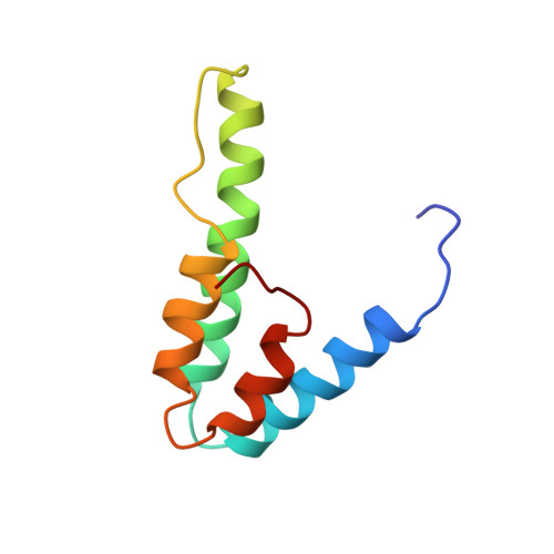
    Gene-specific targeting of the Sin3 corepressor complex by DNA-bound repressors is an important mechanism of gene silencing in eukaryotes. The Sin3 corepressor specifically associates with a diverse group of transcriptional repressors, including members of the Mad family, that play crucial roles in development. The NMR structure of the complex formed by the PAH2 domain of mammalian Sin3A with the transrepression domain (SID) of human Mad1 reveals that both domains undergo mutual folding transitions upon complex formation generating an unusual left-handed four-helix bundle structure and an amphipathic alpha helix, respectively. The SID helix is wedged within a deep hydrophobic pocket defined by two PAH2 helices. Structure-function analyses of the Mad-Sin3 complex provide a basis for understanding the underlying mechanism(s) that lead to gene silencing.


  • Organizational Affiliation
    • Department of Biochemistry, Molecular Biology, and Cell Biology, Northwestern University, Evanston, IL 60208, USA.

Macromolecule Content 

  • Total Structure Weight: 12.31 kDa 
  • Atom Count: 869 
  • Modeled Residue Count: 105 
  • Deposited Residue Count: 105 
  • Unique protein chains: 2

Macromolecules

Find similar proteins by:  Sequence   |   3D Structure  
Entity ID: 1
MoleculeChains  Sequence LengthOrganismDetailsImage
MAD1 PROTEIN16N/AMutation(s): 0 
UniProt & NIH Common Fund Data Resources
Find proteins for Q05195 (Homo sapiens)
Explore Q05195 
Go to UniProtKB:  Q05195
PHAROS:  Q05195
GTEx:  ENSG00000059728 
Entity Groups
Sequence Clusters30% Identity50% Identity70% Identity90% Identity95% Identity100% Identity
UniProt GroupQ05195
Sequence Annotations
Expand
Reference Sequence
Find similar proteins by:|  3D Structure
Entity ID: 2
MoleculeChains  Sequence LengthOrganismDetailsImage
SIN3A89Mus musculusMutation(s): 0 
UniProt
Find proteins for Q60520 (Mus musculus)
Explore Q60520 
Go to UniProtKB:  Q60520
Entity Groups
Sequence Clusters30% Identity50% Identity70% Identity90% Identity95% Identity100% Identity
UniProt GroupQ60520
Sequence Annotations
Expand
Reference Sequence

Experimental Data & Validation

Experimental Data

  • Method: SOLUTION NMR
  • Conformers Calculated: 25 
  • Conformers Submitted: 15 
  • Selection Criteria: structures with favorable non-bond energy,structures with the least restraint violations 

Structure Validation

View Full Validation Report


Entry History 

Deposition Data

Revision History  (Full details and data files)

  • Version 1.0: 2000-12-06
    Type: Initial release
  • Version 1.1: 2008-04-27
    Changes: Version format compliance
  • Version 1.2: 2011-07-13
    Changes: Version format compliance
  • Version 1.3: 2022-02-23
    Changes: Data collection, Database references, Derived calculations
  • Version 1.4: 2024-05-22
    Changes: Data collection