1G0Y

IL-1 RECEPTOR TYPE 1 COMPLEXED WITH ANTAGONIST PEPTIDE AF10847


Experimental Data Snapshot

  • Method: X-RAY DIFFRACTION
  • Resolution: 3.00 Å
  • R-Value Free: 0.332 
  • R-Value Work: 0.223 

wwPDB Validation   3D Report Full Report


This is version 1.4 of the entry. See complete history


Literature

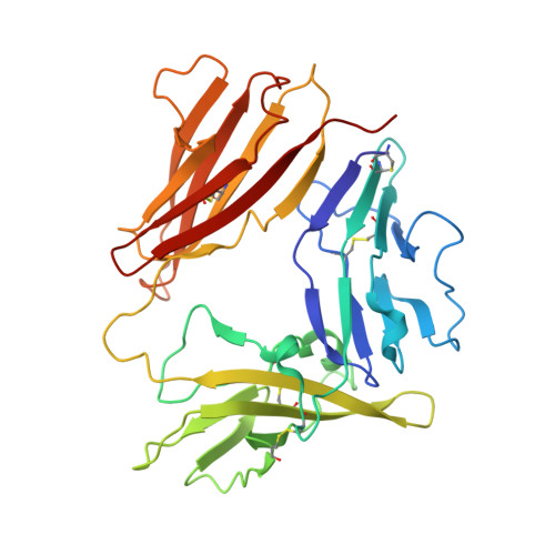
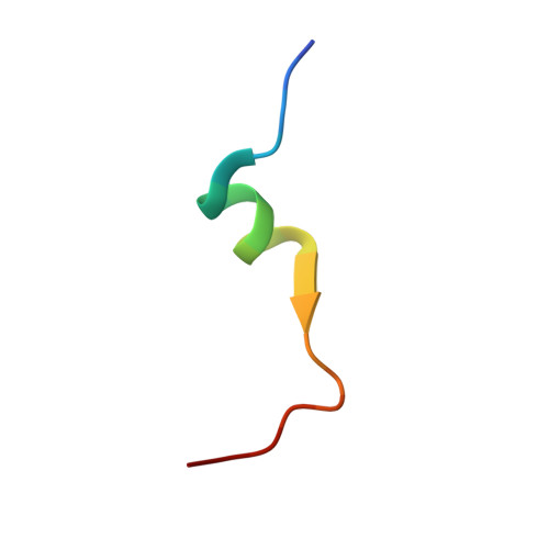
X-ray crystal structure of a small antagonist peptide bound to interleukin-1 receptor type 1.

Vigers, G.P.Dripps, D.J.Edwards III, C.K.Brandhuber, B.J.

(2000) J Biol Chem 275: 36927-36933

  • DOI: https://doi.org/10.1074/jbc.M006071200
  • Primary Citation of Related Structures:  
    1G0Y

  • PubMed Abstract: 

    Interleukin (IL-1)alpha and IL-1beta are important mediators of inflammation. The binding of IL-1 to interleukin-1 receptor (IL-1R) type 1 is the initial step in IL-1 signal transduction and therefore is a tempting target for anti-inflammatory therapeutics. To advance our understanding of IL-1R1 binding interactions, we have determined the structure of the extracellular domains of IL-1R1 bound to a 21-amino acid IL-1 antagonist peptide at 3.0-A resolution. The antagonist peptide binds to the domain 1/2 junction of the receptor, which is a conserved binding site for IL-1beta and IL-1 receptor antagonist (IL-1ra). This co-crystal structure also reveals that considerable flexibility is present in IL-1R1 because the carboxyl-terminal domain of the receptor is rotated almost 170 degrees relative to the first two domains of the receptor compared with the previously solved IL-1R1.ligand structures. The structure shows an unexpected binding mode for the peptide and may contribute to the design of smaller IL-1R antagonists.


  • Organizational Affiliation

    Department of Inflammation Research, Amgen, Incorporated, Boulder, Colorado 80301, USA. gvigers@arraybiopharma.com


Macromolecules
Find similar proteins by:  (by identity cutoff)  |  3D Structure
Entity ID: 1
MoleculeChains Sequence LengthOrganismDetailsImage
INTERLEUKIN-1 RECEPTOR, TYPE IA [auth R]312Homo sapiensMutation(s): 0 
UniProt & NIH Common Fund Data Resources
Find proteins for P14778 (Homo sapiens)
Explore P14778 
Go to UniProtKB:  P14778
PHAROS:  P14778
GTEx:  ENSG00000115594 
Entity Groups  
Sequence Clusters30% Identity50% Identity70% Identity90% Identity95% Identity100% Identity
UniProt GroupP14778
Sequence Annotations
Expand
  • Reference Sequence

Find similar proteins by:  Sequence   |   3D Structure  

Entity ID: 2
MoleculeChains Sequence LengthOrganismDetailsImage
ANTAGONIST PEPTIDE AF10847B [auth I]21N/AMutation(s): 0 
Entity Groups  
Sequence Clusters30% Identity50% Identity70% Identity90% Identity95% Identity100% Identity
Sequence Annotations
Expand
  • Reference Sequence
Experimental Data & Validation

Experimental Data

  • Method: X-RAY DIFFRACTION
  • Resolution: 3.00 Å
  • R-Value Free: 0.332 
  • R-Value Work: 0.223 
  • Space Group: P 65 2 2
Unit Cell:
Length ( Å )Angle ( ˚ )
a = 95.898α = 90
b = 95.898β = 90
c = 208.017γ = 120
Software Package:
Software NamePurpose
DENZOdata reduction
SCALEPACKdata scaling
MLPHAREphasing
X-PLORrefinement

Structure Validation

View Full Validation Report


Entry History 

Deposition Data

Revision History  (Full details and data files)

  • Version 1.0: 2000-10-25
    Type: Initial release
  • Version 1.1: 2008-04-27
    Changes: Version format compliance
  • Version 1.2: 2011-07-13
    Changes: Version format compliance
  • Version 1.3: 2018-01-24
    Changes: Database references
  • Version 1.4: 2018-01-31
    Changes: Database references, Experimental preparation