1FZA

CRYSTAL STRUCTURE OF FIBRINOGEN FRAGMENT D


Experimental Data Snapshot

  • Method: X-RAY DIFFRACTION
  • Resolution: 2.90 Å
  • R-Value Free: 0.363 
  • R-Value Work: 0.263 
  • R-Value Observed: 0.263 

wwPDB Validation   3D Report Full Report


This is version 1.3 of the entry. See complete history


Literature

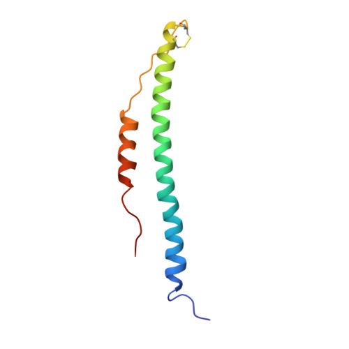
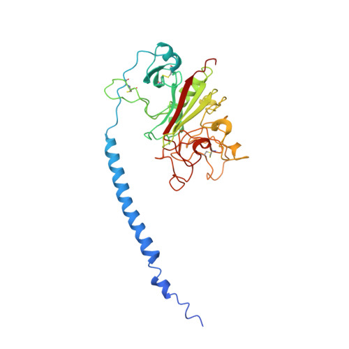
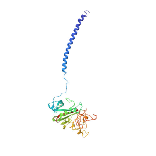
Crystal structures of fragment D from human fibrinogen and its crosslinked counterpart from fibrin.

Spraggon, G.Everse, S.J.Doolittle, R.F.

(1997) Nature 389: 455-462

  • DOI: https://doi.org/10.1038/38947
  • Primary Citation of Related Structures:  
    1FZA, 1FZB

  • PubMed Abstract: 
  • In blood coagulation, units of the protein fibrinogen pack together to form a fibrin clot, but a crystal structure for fibrinogen is needed to understand how this is achieved. The structure of a core fragment (fragment D) from human fibrinogen has now been determined to 2 ...

    In blood coagulation, units of the protein fibrinogen pack together to form a fibrin clot, but a crystal structure for fibrinogen is needed to understand how this is achieved. The structure of a core fragment (fragment D) from human fibrinogen has now been determined to 2.9 A resolution. The 86K three-chained structure consists of a coiled-coil region and two homologous globular entitles oriented at approximately 130 degrees to each other. Additionally, the covalently bound dimer of fragment D, known as 'double-D', was isolated from human fibrin, crystallized in the presence of a Gly-Pro-Arg-Pro-amide peptide ligand, which simulates the donor polymerization site, and its structure solved by molecular replacement with the model of fragment D.


    Organizational Affiliation

    Center for Molecular Genetics, University of California, San Diego, La Jolla 92093-0634, USA.


Macromolecules
Find similar proteins by:  (by identity cutoff)  |  3D Structure
Entity ID: 1
MoleculeChainsSequence LengthOrganismDetailsImage
FIBRINOGEN
A, D
87Homo sapiensMutation(s): 0 
Gene Names: FGA
UniProt & NIH Common Fund Data Resources
Find proteins for P02671 (Homo sapiens)
Explore P02671 
Go to UniProtKB:  P02671
PHAROS:  P02671
Entity Groups  
Sequence Clusters30% Identity50% Identity70% Identity90% Identity95% Identity100% Identity
UniProt GroupP02671
Protein Feature View
Expand
  • Reference Sequence
Find similar proteins by:  (by identity cutoff)  |  3D Structure
Entity ID: 2
MoleculeChainsSequence LengthOrganismDetailsImage
FIBRINOGEN
B, E
328Homo sapiensMutation(s): 0 
Gene Names: FGB
UniProt & NIH Common Fund Data Resources
Find proteins for P02675 (Homo sapiens)
Explore P02675 
Go to UniProtKB:  P02675
PHAROS:  P02675
Entity Groups  
Sequence Clusters30% Identity50% Identity70% Identity90% Identity95% Identity100% Identity
UniProt GroupP02675
Protein Feature View
Expand
  • Reference Sequence
Find similar proteins by:  (by identity cutoff)  |  3D Structure
Entity ID: 3
MoleculeChainsSequence LengthOrganismDetailsImage
FIBRINOGEN
C, F
319Homo sapiensMutation(s): 0 
Gene Names: FGGPRO2061
UniProt & NIH Common Fund Data Resources
Find proteins for P02679 (Homo sapiens)
Explore P02679 
Go to UniProtKB:  P02679
PHAROS:  P02679
Entity Groups  
Sequence Clusters30% Identity50% Identity70% Identity90% Identity95% Identity100% Identity
UniProt GroupP02679
Protein Feature View
Expand
  • Reference Sequence
Small Molecules
Ligands 2 Unique
IDChainsName / Formula / InChI Key2D Diagram3D Interactions
NAG
Query on NAG

Download Ideal Coordinates CCD File 
G [auth B],
I [auth E]
2-acetamido-2-deoxy-beta-D-glucopyranose
C8 H15 N O6
OVRNDRQMDRJTHS-FMDGEEDCSA-N
 Ligand Interaction
CA
Query on CA

Download Ideal Coordinates CCD File 
H [auth C],
J [auth F]
CALCIUM ION
Ca
BHPQYMZQTOCNFJ-UHFFFAOYSA-N
 Ligand Interaction
Experimental Data & Validation

Experimental Data

  • Method: X-RAY DIFFRACTION
  • Resolution: 2.90 Å
  • R-Value Free: 0.363 
  • R-Value Work: 0.263 
  • R-Value Observed: 0.263 
  • Space Group: P 1 21 1
Unit Cell:
Length ( Å )Angle ( ˚ )
a = 107.72α = 90
b = 48.08β = 105.7
c = 167.56γ = 90
Software Package:
Software NamePurpose
PHASESphasing
X-PLORrefinement

Structure Validation

View Full Validation Report


Entry History 

Deposition Data

Revision History  (Full details and data files)

  • Version 1.0: 1997-12-03
    Type: Initial release
  • Version 1.1: 2008-03-24
    Changes: Version format compliance
  • Version 1.2: 2011-07-13
    Changes: Non-polymer description, Version format compliance
  • Version 1.3: 2020-07-29
    Type: Remediation
    Reason: Carbohydrate remediation
    Changes: Advisory, Data collection, Database references, Derived calculations, Structure summary