1FXV

PENICILLIN ACYLASE MUTANT IMPAIRED IN CATALYSIS WITH PENICILLIN G IN THE ACTIVE SITE


Experimental Data Snapshot

  • Method: X-RAY DIFFRACTION
  • Resolution: 2.25 Å
  • R-Value Free: 0.236 
  • R-Value Work: 0.187 

wwPDB Validation   3D Report Full Report


Ligand Structure Quality Assessment 


This is version 1.5 of the entry. See complete history


Literature

Characterization of the beta-lactam binding site of penicillin acylase of Escherichia coli by structural and site-directed mutagenesis studies.

Alkema, W.B.Hensgens, C.M.Kroezinga, E.H.de Vries, E.Floris, R.van der Laan, J.M.Dijkstra, B.W.Janssen, D.B.

(2000) Protein Eng 13: 857-863

  • DOI: https://doi.org/10.1093/protein/13.12.857
  • Primary Citation of Related Structures:  
    1FXH, 1FXV

  • PubMed Abstract: 

    The binding of penicillin to penicillin acylase was studied by X-ray crystallography. The structure of the enzyme-substrate complex was determined after soaking crystals of an inactive betaN241A penicillin acylase mutant with penicillin G. Binding of the substrate induces a conformational change, in which the side chains of alphaF146 and alphaR145 move away from the active site, which allows the enzyme to accommodate penicillin G. In the resulting structure, the beta-lactam binding site is formed by the side chains of alphaF146 and betaF71, which have van der Waals interactions with the thiazolidine ring of penicillin G and the side chain of alphaR145 that is connected to the carboxylate group of the ligand by means of hydrogen bonding via two water molecules. The backbone oxygen of betaQ23 forms a hydrogen bond with the carbonyl oxygen of the phenylacetic acid moiety through a bridging water molecule. Kinetic studies revealed that the site-directed mutants alphaF146Y, alphaF146A and alphaF146L all show significant changes in their interaction with the beta-lactam substrates as compared with the wild type. The alphaF146Y mutant had the same affinity for 6-aminopenicillanic acid as the wild-type enzyme, but was not able to synthesize penicillin G from phenylacetamide and 6-aminopenicillanic acid. The alphaF146L and alphaF146A enzymes had a 3-5-fold decreased affinity for 6-aminopenicillanic acid, but synthesized penicillin G more efficiently than the wild type. The combined results of the structural and kinetic studies show the importance of alphaF146 in the beta-lactam binding site and provide leads for engineering mutants with improved synthetic properties.


  • Organizational Affiliation

    Department of Biochemistry, BIOSON Research Institute, Groningen Biomolecular Sciences and Biotechnology Institute, University of Groningen, Nijenborgh 4, 9747 AG Groningen, The Netherlands.


Macromolecules
Find similar proteins by:  (by identity cutoff)  |  3D Structure
Entity ID: 1
MoleculeChains Sequence LengthOrganismDetailsImage
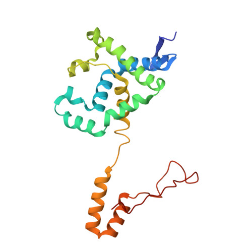
PENICILLIN ACYLASE209Escherichia coliMutation(s): 0 
EC: 3.5.1.11
UniProt
Find proteins for P06875 (Escherichia coli)
Explore P06875 
Go to UniProtKB:  P06875
Entity Groups  
Sequence Clusters30% Identity50% Identity70% Identity90% Identity95% Identity100% Identity
UniProt GroupP06875
Sequence Annotations
Expand
  • Reference Sequence
Find similar proteins by:  (by identity cutoff)  |  3D Structure
Entity ID: 2
MoleculeChains Sequence LengthOrganismDetailsImage
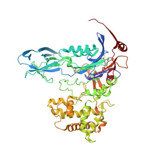
PENICILLIN ACYLASE557Escherichia coliMutation(s): 2 
EC: 3.5.1.11
UniProt
Find proteins for P06875 (Escherichia coli)
Explore P06875 
Go to UniProtKB:  P06875
Entity Groups  
Sequence Clusters30% Identity50% Identity70% Identity90% Identity95% Identity100% Identity
UniProt GroupP06875
Sequence Annotations
Expand
  • Reference Sequence
Small Molecules
Ligands 2 Unique
IDChains Name / Formula / InChI Key2D Diagram3D Interactions
PNN
Query on PNN

Download Ideal Coordinates CCD File 
C [auth B]PENICILLIN G
C16 H18 N2 O4 S
JGSARLDLIJGVTE-MBNYWOFBSA-N
CA
Query on CA

Download Ideal Coordinates CCD File 
D [auth B]CALCIUM ION
Ca
BHPQYMZQTOCNFJ-UHFFFAOYSA-N
Experimental Data & Validation

Experimental Data

  • Method: X-RAY DIFFRACTION
  • Resolution: 2.25 Å
  • R-Value Free: 0.236 
  • R-Value Work: 0.187 
  • Space Group: P 1
Unit Cell:
Length ( Å )Angle ( ˚ )
a = 50.93α = 72.85
b = 63.966β = 73.77
c = 64.189γ = 74.06
Software Package:
Software NamePurpose
CNSrefinement
DENZOdata reduction
SCALEPACKdata scaling
CNSphasing

Structure Validation

View Full Validation Report


Ligand Structure Quality Assessment 


Entry History 

Deposition Data

Revision History  (Full details and data files)

  • Version 1.0: 2001-03-28
    Type: Initial release
  • Version 1.1: 2008-04-27
    Changes: Version format compliance
  • Version 1.2: 2011-07-13
    Changes: Version format compliance
  • Version 1.3: 2017-02-08
    Changes: Structure summary
  • Version 1.4: 2021-11-03
    Changes: Database references, Derived calculations
  • Version 1.5: 2024-02-07
    Changes: Data collection