1FX5

CRYSTAL STRUCTURE ANALYSIS OF ULEX EUROPAEUS LECTIN I


Experimental Data Snapshot

  • Method: X-RAY DIFFRACTION
  • Resolution: 2.20 Å
  • R-Value Free: 0.217 
  • R-Value Work: 0.171 
  • R-Value Observed: 0.171 

wwPDB Validation   3D Report Full Report


This is version 2.1 of the entry. See complete history


Literature

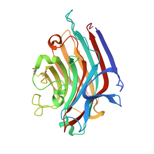
The 2.2 A resolution structure of the O(H) blood-group-specific lectin I from Ulex europaeus.

Audette, G.F.Vandonselaar, M.Delbaere, L.T.

(2000) J Mol Biol 304: 423-433

  • DOI: https://doi.org/10.1006/jmbi.2000.4214
  • Primary Citation of Related Structures:  
    1FX5

  • PubMed Abstract: 

    The tertiary and quaternary structure of the lectin I from Ulex europaeus (UE-I) has been determined to 2.2 A resolution. UE-I is a dimeric metalloglycoprotein that binds the H-type 2 human blood group determinant [alpha-L-Fucalpha(1-->2)-beta-D-Galbeta(1-->4)-beta-D-Glc NAcalpha-]. Nine changes from the published amino acid sequence were necessary to account for the electron density. The quaternary structural organization of UE-I is that of the most commonly occurring legume lectin dimer. The tertiary structure of the monomeric subunits is similar to that in the conventional lectin subunit; however, some structural differences are noted. These differences include a four-stranded anti-parallel "S" sheet in UE-I versus the five-stranded S sheet in other lectin monomers. The Ala residue of the Ala-Asp cis-peptide bond present in the carbohydrate-binding site of the conventional lectin monomer is replaced with a Thr in the UE-I structure. Also, a novel disulfide bridge linking Cys115 and Cys150 is present. There are two metallic ions, one calcium and the other manganese, per subunit. N-linked oligosaccharides are at residues 23 and 111 of each subunit. One molecule of R-2-methyl-2, 4-pentanediol (R-MPD) is present in a shallow depression on the surface of each subunit. In order to examine the binding of the H-type 2 blood group determinant by UE-I, its beta-methyl glycoside (H-type 2-OMe) was docked into the binding site of R-MPD. The epitope previously identified for H-type 2-OMe by chemical mapping proved, with only minor adjustment of amino acid residues, to be complementary to the shallow cavity occupied by R-MPD in the structure. Several key interactions have been proposed between the H-type 2-OMe and UE-I.


  • Organizational Affiliation

    Department of Biochemistry, University of Saskatchewan, 107 Wiggins Road, Saskatoon, Saskatchewan, S7N 5E5, Canada.


Macromolecules
Find similar proteins by:  (by identity cutoff)  |  3D Structure
Entity ID: 1
MoleculeChains Sequence LengthOrganismDetailsImage
ANTI-H(O) LECTIN I
A, B
242Ulex europaeusMutation(s): 0 
UniProt
Find proteins for P22972 (Ulex europaeus)
Explore P22972 
Go to UniProtKB:  P22972
Entity Groups  
Sequence Clusters30% Identity50% Identity70% Identity90% Identity95% Identity100% Identity
UniProt GroupP22972
Sequence Annotations
Expand
  • Reference Sequence
Oligosaccharides

Help

Entity ID: 2
MoleculeChains Length2D Diagram Glycosylation3D Interactions
Xylitol-(1-2)-[alpha-D-mannopyranose-(1-3)][alpha-D-mannopyranose-(1-6)]beta-D-mannopyranose-(1-4)-2-acetamido-2-deoxy-beta-D-glucopyranose-(1-4)-[alpha-L-fucopyranose-(1-3)]2-acetamido-2-deoxy-beta-D-glucopyranose
C
7N/AN-Glycosylation
Entity ID: 3
MoleculeChains Length2D Diagram Glycosylation3D Interactions
alpha-L-fucopyranose-(1-3)-[2-acetamido-2-deoxy-beta-D-glucopyranose-(1-4)]2-acetamido-2-deoxy-beta-D-glucopyranose
D, E
3N-Glycosylation
Experimental Data & Validation

Experimental Data

  • Method: X-RAY DIFFRACTION
  • Resolution: 2.20 Å
  • R-Value Free: 0.217 
  • R-Value Work: 0.171 
  • R-Value Observed: 0.171 
  • Space Group: C 1 2 1
Unit Cell:
Length ( Å )Angle ( ˚ )
a = 79.61α = 90
b = 70.67β = 108.6
c = 122.15γ = 90
Software Package:
Software NamePurpose
MADNESSdata collection
TRUNCATEdata reduction
AMoREphasing
X-PLORrefinement
MADNESSdata reduction
CCP4data scaling

Structure Validation

View Full Validation Report


Entry History 

Deposition Data

Revision History  (Full details and data files)

  • Version 1.0: 2001-01-17
    Type: Initial release
  • Version 1.1: 2008-04-27
    Changes: Version format compliance
  • Version 1.2: 2011-07-13
    Changes: Non-polymer description, Version format compliance
  • Version 1.3: 2017-10-04
    Changes: Refinement description
  • Version 1.4: 2018-04-18
    Changes: Data collection
  • Version 2.0: 2020-07-29
    Type: Remediation
    Reason: Carbohydrate remediation
    Changes: Atomic model, Data collection, Database references, Derived calculations, Structure summary
  • Version 2.1: 2023-08-09
    Changes: Data collection, Database references, Refinement description, Structure summary