1FVF

CRYSTAL STRUCTURE ANALYSIS OF NEURONAL SEC1 FROM THE SQUID L. PEALEI


Experimental Data Snapshot

  • Method: X-RAY DIFFRACTION
  • Resolution: 3.20 Å
  • R-Value Free: 0.288 
  • R-Value Work: 0.243 
  • R-Value Observed: 0.245 

wwPDB Validation   3D Report Full Report


This is version 1.4 of the entry. See complete history


Literature

Crystal structures of neuronal squid Sec1 implicate inter-domain hinge movement in the release of t-SNAREs.

Bracher, A.Weissenhorn, W.

(2001) J Mol Biol 306: 7-13

  • DOI: https://doi.org/10.1006/jmbi.2000.4347
  • Primary Citation of Related Structures:  
    1FVF, 1FVH

  • PubMed Abstract: 

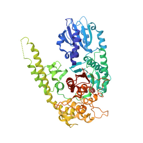
    Sec1 molecules associate with t-SNAREs from the syntaxin family in a heterodimeric complex that plays an essential role in vesicle transport and membrane fusion. Neuronal rat n-Sec1 has an arch-shaped three-domain structure, which binds syntaxin 1a through contacts in domains 1 and 3. In both rat nSec1 and homologous squid s-Sec1, a potential effector-molecule binding-pocket is shaped by residues from domains 1 and 2 and is localized on the opposite side of the syntaxin 1a interaction site. Comparison of several crystal forms of unliganded neuronal squid Sec1 indicates a hinge region between domains 1 and 2 which allows domain 1 to rotate along a central axis. This movement could release syntaxin 1a upon interaction with a yet unspecified Sec1 effector molecule(s). The binding of an effector protein may also directly affect the conformation of the helical hairpin of domain 3, which contributes the other significant syntaxin 1a binding sites in the rat nSec1/syntaxin 1a complex structure but adopts multiple conformations in the unliganded s-Sec1 structures reported here.


  • Organizational Affiliation

    European Molecular Biology Laboratory (EMBL), 6 rue Jules Horowitz, Grenoble, 38000, France.


Macromolecules
Find similar proteins by:  (by identity cutoff)  |  3D Structure
Entity ID: 1
MoleculeChains Sequence LengthOrganismDetailsImage
SEC1
A, B
591Doryteuthis pealeiiMutation(s): 0 
UniProt
Find proteins for O62547 (Doryteuthis pealeii)
Explore O62547 
Go to UniProtKB:  O62547
Entity Groups  
Sequence Clusters30% Identity50% Identity70% Identity90% Identity95% Identity100% Identity
UniProt GroupO62547
Sequence Annotations
Expand
  • Reference Sequence
Experimental Data & Validation

Experimental Data

  • Method: X-RAY DIFFRACTION
  • Resolution: 3.20 Å
  • R-Value Free: 0.288 
  • R-Value Work: 0.243 
  • R-Value Observed: 0.245 
  • Space Group: P 31 2 1
Unit Cell:
Length ( Å )Angle ( ˚ )
a = 118.517α = 90
b = 118.517β = 90
c = 192.621γ = 120
Software Package:
Software NamePurpose
AMoREphasing
CNSrefinement
DENZOdata reduction
SCALEPACKdata scaling

Structure Validation

View Full Validation Report


Entry History 

Deposition Data

Revision History  (Full details and data files)

  • Version 1.0: 2001-01-31
    Type: Initial release
  • Version 1.1: 2008-04-27
    Changes: Version format compliance
  • Version 1.2: 2011-07-13
    Changes: Version format compliance
  • Version 1.3: 2011-07-27
    Changes: Atomic model, Derived calculations
  • Version 1.4: 2023-08-09
    Changes: Data collection, Database references, Refinement description