1FUY

CRYSTAL STRUCTURE OF BETAA169L/BETAC170W DOUBLE MUTANT OF TRYPTOPHAN SYNTHASE COMPLEXED WITH 5-FLUORO-INDOLE-PROPANOL PHOSPHATE


Experimental Data Snapshot

  • Method: X-RAY DIFFRACTION
  • Resolution: 2.25 Å
  • R-Value Free: 0.224 
  • R-Value Work: 0.170 
  • R-Value Observed: 0.178 

wwPDB Validation   3D Report Full Report


Ligand Structure Quality Assessment 


This is version 1.4 of the entry. See complete history


Literature

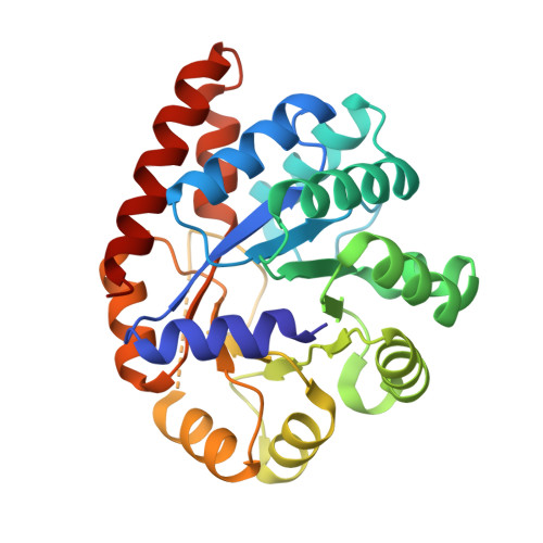
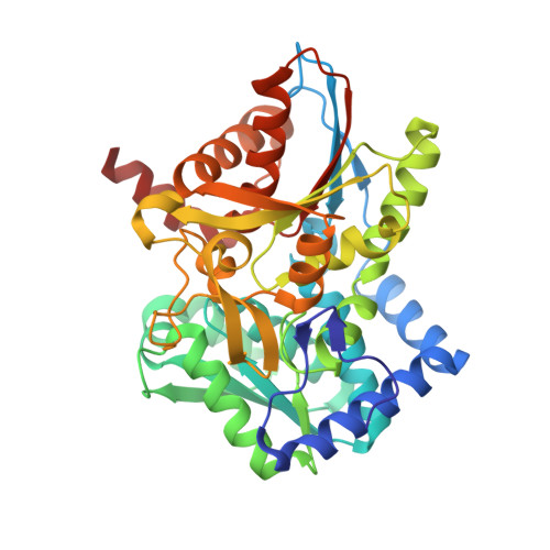
Structural basis for the impaired channeling and allosteric inter-subunit communication in the beta A169L/beta C170W mutant of tryptophan synthase.

Weyand, M.Schlichting, I.

(2000) J Biol Chem 275: 41058-41063

  • DOI: https://doi.org/10.1074/jbc.C000479200
  • Primary Citation of Related Structures:  
    1FUY

  • PubMed Abstract: 

    We determined the 2.25 A resolution crystal structure of the betaA169L/betaC170W mutant form of the tryptophan synthase alpha(2)beta(2) complex from Salmonella typhimurium complexed with the alpha-active site substrate analogue 5-fluoro-indole-propanol-phosphate to identify the structural basis for the changed kinetic properties of the mutant (Anderson, K. S., Kim, A. Y., Quillen, J. M., Sayers, E., Yang, X. J., and Miles, E. W. (1995) J. Biol. Chem. 270, 29936-29944). Comparison with the wild-type enzyme showed that the betaTrp(170) side chain occludes the tunnel connecting the alpha- and beta-active sites, explaining the accumulation of the intermediate indole during a single enzyme turnover. To prevent a steric clash between betaLeu(169) and betaGly(135), located in the beta-sheet of the COMM (communication) domain (betaGly(102)-betaGly(189)), the latter reorganizes. The changed COMM domain conformation results in a loss of the hydrogen bonding networks between the alpha- and beta-active sites, explaining the poor activation of the alpha-reaction upon formation of the aminoacrylate complex at the beta-active site. The 100-fold reduced affinity for serine seems to result from a movement of betaAsp(305) away from the beta-active site so that it cannot interact with the hydroxyl group of a pyridoxal phosphate-bound serine. The proposed structural dissection of the effects of each single mutation in the betaA169L/betaC170W mutant would explain the very different kinetics of this mutant and betaC170F.


  • Organizational Affiliation

    Max Planck Institut für Molekulare Physiologie, Abteilung Physikalische Biochemie, Otto-Hahn-Strabetae 11, 44227 Dortmund, Germany.


Macromolecules
Find similar proteins by:  (by identity cutoff)  |  3D Structure
Entity ID: 1
MoleculeChains Sequence LengthOrganismDetailsImage
TRYPTOPHAN SYNTHASE ALPHA CHAIN268Salmonella enterica subsp. enterica serovar TyphimuriumMutation(s): 0 
Gene Names: TRPA/TRPB
EC: 4.2.1.20
UniProt
Find proteins for P00929 (Salmonella typhimurium (strain LT2 / SGSC1412 / ATCC 700720))
Explore P00929 
Go to UniProtKB:  P00929
Entity Groups  
Sequence Clusters30% Identity50% Identity70% Identity90% Identity95% Identity100% Identity
UniProt GroupP00929
Sequence Annotations
Expand
  • Reference Sequence
Find similar proteins by:  (by identity cutoff)  |  3D Structure
Entity ID: 2
MoleculeChains Sequence LengthOrganismDetailsImage
TRYPTOPHAN SYNTHASE BETA CHAIN396Salmonella enterica subsp. enterica serovar TyphimuriumMutation(s): 2 
Gene Names: TRPA/TRPB
EC: 4.2.1.20
UniProt
Find proteins for P0A2K1 (Salmonella typhimurium (strain LT2 / SGSC1412 / ATCC 700720))
Explore P0A2K1 
Go to UniProtKB:  P0A2K1
Entity Groups  
Sequence Clusters30% Identity50% Identity70% Identity90% Identity95% Identity100% Identity
UniProt GroupP0A2K1
Sequence Annotations
Expand
  • Reference Sequence
Small Molecules
Experimental Data & Validation

Experimental Data

  • Method: X-RAY DIFFRACTION
  • Resolution: 2.25 Å
  • R-Value Free: 0.224 
  • R-Value Work: 0.170 
  • R-Value Observed: 0.178 
  • Space Group: C 1 2 1
Unit Cell:
Length ( Å )Angle ( ˚ )
a = 184α = 90
b = 60.7β = 95.75
c = 67.4γ = 90
Software Package:
Software NamePurpose
X-PLORmodel building
REFMACrefinement
DENZOdata reduction
SCALEPACKdata scaling
X-PLORphasing

Structure Validation

View Full Validation Report


Ligand Structure Quality Assessment 


Entry History 

Deposition Data

Revision History  (Full details and data files)

  • Version 1.0: 2001-01-17
    Type: Initial release
  • Version 1.1: 2008-04-27
    Changes: Version format compliance
  • Version 1.2: 2011-07-13
    Changes: Non-polymer description, Version format compliance
  • Version 1.3: 2021-07-21
    Changes: Database references, Derived calculations, Refinement description
  • Version 1.4: 2021-11-03
    Changes: Database references