1FUJ | pdb_00001fuj

PR3 (MYELOBLASTIN)


Experimental Data Snapshot

  • Method: X-RAY DIFFRACTION
  • Resolution: 2.20 Å
  • R-Value Work: 
    0.179 (DCC) 

wwPDB Validation 3D Report Full Report

Validation slider image for 1FUJ

This is version 2.1 of the entry. See complete history

Literature

The crystal structure of PR3, a neutrophil serine proteinase antigen of Wegener's granulomatosis antibodies.

Fujinaga, M.Chernaia, M.M.Halenbeck, R.Koths, K.James, M.N.

(1996) J Mol Biology 261: 267-278

  • DOI: https://doi.org/10.1006/jmbi.1996.0458
  • Primary Citation Related Structures: 
    1FUJ

  • PubMed Abstract: 

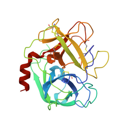
    The crystal structure of PR3, a serine proteinase from the azurophilic granules of human polymorphonuclear neutrophils, has been solved by molecular replacement using the human leukocyte elastase structure. The PR3 structure has been refined to an R-factor (= sigma parallel Fo magnitude of-Fc parallel/sigma magnitude of Fo) of 0.201 for all data in the range of 10.0 to 2.2 A resolution. The enzyme was crystallized in space group P21 with four molecules in the asymmetric unit (Vm approximately equal to 2.6 A/Da). The overall fold consists of two domains of beta-barrel structures typical of the chymotrypsin family of serine proteinases. In general, the substrate binding sites, S4 to S3', are more polar than comparable sites in the related proteinase, human leukocyte elastase. The experimentally observed preference of PR3 for small aliphatic residues at the P1 position of a substrate is explained by the Val to Ile substitution at position 190 when compared to the elastase structure. The substitution of Ala by Asp at position 213 at the back of S1 should not affect its specificity greatly, as the Asp side-chain points back into the interior of the protein. The PR3 structure includes a disaccharide unit (N-linked 2-acetamido-2-deoxy-beta-D-glucopyranose and 1,6-linked alpha-L-fucopyranose) covalently attached to Asn 159. The linear antigenic sites of PR3 reported to react with Wegener's granulomatosis autoantibodies occur in regions of the three-dimensional structure that may implicate the inactive pro-form of the enzyme in the pathogenesis of the disease.


  • Organizational Affiliation
    • Department of Biochemistry, University of Alberta, Edmonton, Canada.

Macromolecule Content 

  • Total Structure Weight: 98.61 kDa 
  • Atom Count: 7,174 
  • Modeled Residue Count: 884 
  • Deposited Residue Count: 884 
  • Unique protein chains: 1

Macromolecules

Find similar proteins by:|  3D Structure
Entity ID: 1
MoleculeChains  Sequence LengthOrganismDetailsImage
PR3
A, B, C, D
221Homo sapiensMutation(s): 0 
EC: 3.4.21.76
UniProt & NIH Common Fund Data Resources
Find proteins for P24158 (Homo sapiens)
Explore P24158 
Go to UniProtKB:  P24158
PHAROS:  P24158
GTEx:  ENSG00000196415 
Entity Groups
Sequence Clusters30% Identity50% Identity70% Identity90% Identity95% Identity100% Identity
UniProt GroupP24158
Glycosylation
Glycosylation Sites: 1Go to GlyGen: P24158-1
Sequence Annotations
Expand
Reference Sequence

Oligosaccharides

Help  
Entity ID: 2
MoleculeChains Length2D Diagram GlycosylationD Interactions
alpha-L-fucopyranose-(1-6)-2-acetamido-2-deoxy-beta-D-glucopyranose
E, F, G, H
2N-Glycosylation
Glycosylation Resources
GlyTouCan: G86851RC
GlyCosmos: G86851RC
GlyGen: G86851RC

Experimental Data & Validation

Experimental Data

  • Method: X-RAY DIFFRACTION
  • Resolution: 2.20 Å
  • R-Value Work:  0.179 (DCC) 
Space Group: P 1 21 1
Unit Cell:
Length ( Å )Angle ( ˚ )
a = 85.603α = 90
b = 54.07β = 90.69
c = 113.506γ = 90
Software Package:
Software NamePurpose
TNTrefinement

Structure Validation

View Full Validation Report


Entry History 

Deposition Data

Revision History  (Full details and data files)

  • Version 1.0: 1996-07-11
    Type: Initial release
  • Version 1.1: 2008-03-24
    Changes: Version format compliance
  • Version 1.2: 2011-07-13
    Changes: Non-polymer description, Version format compliance
  • Version 2.0: 2020-07-29
    Type: Remediation
    Reason: Carbohydrate remediation
    Changes: Advisory, Atomic model, Data collection, Database references, Derived calculations, Other, Structure summary
  • Version 2.1: 2024-10-23
    Changes: Data collection, Database references, Structure summary