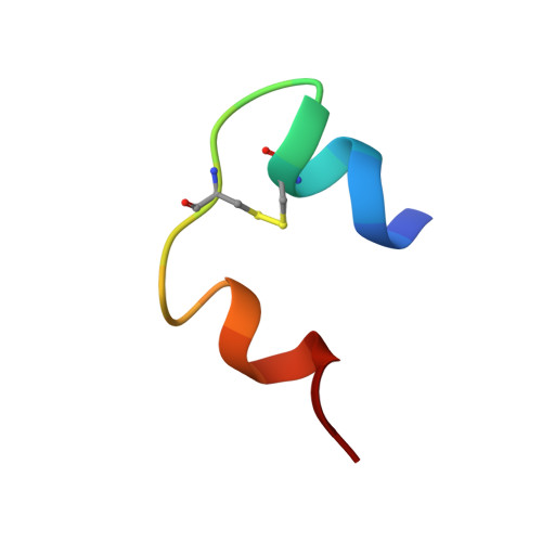
The first protein crystal structure determined from high-resolution X-ray powder diffraction data: a variant of T3R3 human insulin-zinc complex produced by grinding.
Von Dreele, R.B., Stephens, P.W., Smith, G.D., Blessing, R.H.(2000) Acta Crystallogr D Biol Crystallogr 56: 1549-1553
- PubMed: 11092920 Search on PubMed
- DOI: https://doi.org/10.1107/s0907444900013901
- Primary Citation Related Structures:
1FU2, 1FUB - PubMed Abstract:
X-ray diffraction analysis of protein structure is often limited by the availability of suitable crystals. However, the absence of single crystals need not present an insurmountable obstacle in protein crystallography any more than it does in materials science, where powder diffraction techniques have developed to the point where complex oxide, zeolite and small organic molecular structures can often be solved from powder data alone. Here, that fact is demonstrated with the structure solution and refinement of a new variant of the T(3)R(3) Zn-human insulin complex produced by mechanical grinding of a polycrystalline sample. High-resolution synchrotron X-ray powder diffraction data were used to solve this crystal structure by molecular replacement adapted for Rietveld refinement. A complete Rietveld refinement of the 1630-atom protein was achieved by combining 7981 stereochemical restraints with a 4800-step (d(min) = 3.24 A) powder diffraction pattern and yielded the residuals R(wp) = 3.73%, R(p) = 2.84%, R(F)(2) = 8.25%. It was determined that the grinding-induced phase change is accompanied by 9.5 and 17.2 degrees rotations of the two T(3)R(3) complexes that comprise the crystal structure. The material reverts over 2-3 d to recover the original T(3)R(3) crystal structure. A Rietveld refinement of this 815-atom protein by combining 3886 stereochemical restraints with a 6000-step (d(min) = 3.06 A) powder diffraction pattern yielded the residuals R(wp) = 3.46%, R(p) = 2.64%, R(F)(2) = 7.10%. The demonstrated ability to solve and refine a protein crystal structure from powder diffraction data suggests that this approach can be employed, for example, to examine structural changes in a series of protein derivatives in which the structure of one member is known from a single-crystal study.
- Manuel Lujan Jr Neutron Scattering Center, MS H805, Los Alamos National Laboratory, Los Alamos, NM 87545, USA. vondreele@lanl.gov
Organizational Affiliation: