1FQ9

CRYSTAL STRUCTURE OF A TERNARY FGF2-FGFR1-HEPARIN COMPLEX


Experimental Data Snapshot

  • Method: X-RAY DIFFRACTION
  • Resolution: 3.00 Å
  • R-Value Free: 0.282 
  • R-Value Work: 0.229 

wwPDB Validation   3D Report Full Report


This is version 3.0 of the entry. See complete history


Literature

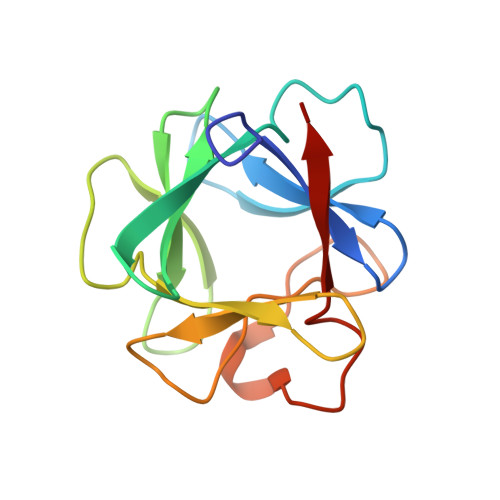
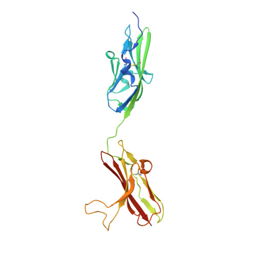
Crystal structure of a ternary FGF-FGFR-heparin complex reveals a dual role for heparin in FGFR binding and dimerization.

Schlessinger, J.Plotnikov, A.N.Ibrahimi, O.A.Eliseenkova, A.V.Yeh, B.K.Yayon, A.Linhardt, R.J.Mohammadi, M.

(2000) Mol Cell 6: 743-750

  • DOI: 10.1016/s1097-2765(00)00073-3
  • Primary Citation of Related Structures:  
    1FQ9

  • PubMed Abstract: 
  • The crystal structure of a dimeric 2:2:2 FGF:FGFR:heparin ternary complex at 3 A resolution has been determined. Within each 1:1 FGF:FGFR complex, heparin makes numerous contacts with both FGF and FGFR, thereby augmenting FGF-FGFR binding. Heparin also interacts with FGFR in the adjoining 1:1 FGF:FGFR complex to promote FGFR dimerization ...

    The crystal structure of a dimeric 2:2:2 FGF:FGFR:heparin ternary complex at 3 A resolution has been determined. Within each 1:1 FGF:FGFR complex, heparin makes numerous contacts with both FGF and FGFR, thereby augmenting FGF-FGFR binding. Heparin also interacts with FGFR in the adjoining 1:1 FGF:FGFR complex to promote FGFR dimerization. The 6-O-sulfate group of heparin plays a pivotal role in mediating both interactions. The unexpected stoichiometry of heparin binding in the structure led us to propose a revised model for FGFR dimerization. Biochemical data in support of this model are also presented. This model provides a structural basis for FGFR activation by small molecule heparin analogs and may facilitate the design of heparin mimetics capable of modulating FGF signaling.


    Organizational Affiliation

    Department of Pharmacology, New York University School of Medicine, New York, New York 10016, USA.


Macromolecules
Find similar proteins by:  (by identity cutoff)  |  3D Structure
Entity ID: 1
MoleculeChainsSequence LengthOrganismDetailsImage
FIBROBLAST GROWTH FACTOR 2A, B132Homo sapiensMutation(s): 2 
Gene Names: FGF2FGFB
UniProt & NIH Common Fund Data Resources
Find proteins for P09038 (Homo sapiens)
Explore P09038 
Go to UniProtKB:  P09038
PHAROS:  P09038
Entity Groups  
Sequence Clusters30% Identity50% Identity70% Identity90% Identity95% Identity100% Identity
UniProt GroupP09038
Protein Feature View
Expand
  • Reference Sequence
Find similar proteins by:  (by identity cutoff)  |  3D Structure
Entity ID: 2
MoleculeChainsSequence LengthOrganismDetailsImage
FIBROBLAST GROWTH FACTOR RECEPTOR 1C, D225Homo sapiensMutation(s): 1 
Gene Names: FGFR1BFGFRCEKFGFBRFLGFLT2HBGFR
EC: 2.7.10.1
UniProt & NIH Common Fund Data Resources
Find proteins for P11362 (Homo sapiens)
Explore P11362 
Go to UniProtKB:  P11362
PHAROS:  P11362
Entity Groups  
Sequence Clusters30% Identity50% Identity70% Identity90% Identity95% Identity100% Identity
UniProt GroupP11362
Protein Feature View
Expand
  • Reference Sequence
Oligosaccharides

Help

Entity ID: 3
MoleculeChainsChain Length2D DiagramGlycosylation3D Interactions
4-deoxy-2-O-sulfo-alpha-L-threo-hex-4-enopyranuronic acid-(1-4)-2-deoxy-6-O-sulfo-2-(sulfoamino)-alpha-D-glucopyranose-(1-4)-2-O-sulfo-alpha-L-idopyranuronic acid-(1-4)-2-deoxy-6-O-sulfo-2-(sulfoamino)-alpha-D-glucopyranose-(1-4)-2-O-sulfo-alpha-L-idopyranuronic acid-(1-4)-2-deoxy-6-O-sulfo-2-(sulfoamino)-alpha-D-glucopyranose-(1-4)-2-O-sulfo-beta-L-altropyranuronic acid-(1-4)-2-deoxy-6-O-sulfo-2-(sulfoamino)-alpha-D-glucopyranoseE 8N/AN/A Oligosaccharides Interaction
Entity ID: 4
MoleculeChainsChain Length2D DiagramGlycosylation3D Interactions
4-deoxy-2-O-sulfo-alpha-L-threo-hex-4-enopyranuronic acid-(1-4)-2-deoxy-6-O-sulfo-2-(sulfoamino)-alpha-D-glucopyranose-(1-4)-2-O-sulfo-alpha-L-idopyranuronic acid-(1-4)-2-deoxy-6-O-sulfo-2-(sulfoamino)-alpha-D-glucopyranose-(1-4)-2-O-sulfo-alpha-L-idopyranuronic acid-(1-4)-2-deoxy-6-O-sulfo-2-(sulfoamino)-alpha-D-glucopyranoseF 6N/A Oligosaccharides Interaction
Experimental Data & Validation

Experimental Data

  • Method: X-RAY DIFFRACTION
  • Resolution: 3.00 Å
  • R-Value Free: 0.282 
  • R-Value Work: 0.229 
  • Space Group: P 41 21 2
Unit Cell:
Length ( Å )Angle ( ˚ )
a = 98.888α = 90
b = 98.888β = 90
c = 196.292γ = 90
Software Package:
Software NamePurpose
DENZOdata reduction
SCALEPACKdata scaling
CNSrefinement
CNSphasing

Structure Validation

View Full Validation Report


Entry History 

Deposition Data

Revision History  (Full details and data files)

  • Version 1.0: 2000-09-27
    Type: Initial release
  • Version 1.1: 2008-04-27
    Changes: Version format compliance
  • Version 1.2: 2011-07-13
    Changes: Non-polymer description, Version format compliance
  • Version 2.0: 2020-07-29
    Type: Remediation
    Reason: Carbohydrate remediation
    Changes: Advisory, Atomic model, Data collection, Derived calculations, Structure summary
  • Version 3.0: 2021-11-03
    Changes: Atomic model, Data collection, Database references, Derived calculations, Structure summary