1FOO

BOVINE ENDOTHELIAL NITRIC OXIDE SYNTHASE HEME DOMAIN COMPLEXED WITH L-ARG AND NO(H4B-FREE)


Experimental Data Snapshot

  • Method: X-RAY DIFFRACTION
  • Resolution: 2.00 Å
  • R-Value Free: 0.253 
  • R-Value Work: 0.223 
  • R-Value Observed: 0.223 

wwPDB Validation   3D Report Full Report


Ligand Structure Quality Assessment 


This is version 1.3 of the entry. See complete history


Literature

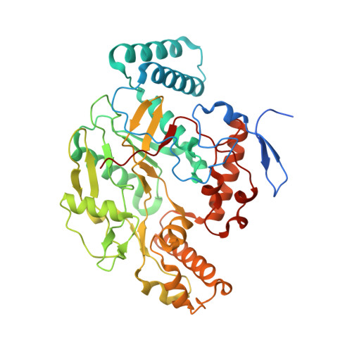
Crystallographic studies on endothelial nitric oxide synthase complexed with nitric oxide and mechanism-based inhibitors.

Li, H.Raman, C.S.Martasek, P.Masters, B.S.Poulos, T.L.

(2001) Biochemistry 40: 5399-5406

  • DOI: https://doi.org/10.1021/bi002658v
  • Primary Citation of Related Structures:  
    1ED6, 1FOI, 1FOL, 1FOO, 1FOP

  • PubMed Abstract: 

    The crystal structure of the endothelial nitric oxide synthase (NOS) heme domain complexed with NO reveals close hydrogen bonding interactions between NO and the terminal guanidino nitrogen of the substrate, L-arginine. Dioxygen is expected to bind in a similar mode which will facilitate proton abstraction from L-Arg to dioxygen, a required step for O-O bond cleavage. Structures of mechanism-based NOS inhibitors, N(5)-(1-iminoethyl)-L-ornithine and N-(3-(aminomethyl)benzyl)acetamidine, provide clues on how this class of compounds operate as suicide substrate inhibitors leading to heme oxidation.


  • Organizational Affiliation

    Departments of Molecular Biology & Biochemistry and Physiology & Biophysics and Program in Macromolecular Structure, University of California, Irvine 92697-3900, USA.


Macromolecules
Find similar proteins by:  (by identity cutoff)  |  3D Structure
Entity ID: 1
MoleculeChains Sequence LengthOrganismDetailsImage
NITRIC-OXIDE SYNTHASE
A, B
444Bos taurusMutation(s): 0 
EC: 1.14.13.39
UniProt
Find proteins for P29473 (Bos taurus)
Explore P29473 
Go to UniProtKB:  P29473
Entity Groups  
Sequence Clusters30% Identity50% Identity70% Identity90% Identity95% Identity100% Identity
UniProt GroupP29473
Sequence Annotations
Expand
  • Reference Sequence
Small Molecules
Ligands 7 Unique
IDChains Name / Formula / InChI Key2D Diagram3D Interactions
HEM
Query on HEM

Download Ideal Coordinates CCD File 
F [auth A],
N [auth B]
PROTOPORPHYRIN IX CONTAINING FE
C34 H32 Fe N4 O4
KABFMIBPWCXCRK-RGGAHWMASA-L
ARG
Query on ARG

Download Ideal Coordinates CCD File 
H [auth A],
P [auth B]
ARGININE
C6 H15 N4 O2
ODKSFYDXXFIFQN-BYPYZUCNSA-O
CAC
Query on CAC

Download Ideal Coordinates CCD File 
C [auth A],
L [auth B]
CACODYLATE ION
C2 H6 As O2
OGGXGZAMXPVRFZ-UHFFFAOYSA-M
GOL
Query on GOL

Download Ideal Coordinates CCD File 
I [auth A],
J [auth A],
K [auth A],
Q [auth B]
GLYCEROL
C3 H8 O3
PEDCQBHIVMGVHV-UHFFFAOYSA-N
ZN
Query on ZN

Download Ideal Coordinates CCD File 
E [auth A]ZINC ION
Zn
PTFCDOFLOPIGGS-UHFFFAOYSA-N
ACT
Query on ACT

Download Ideal Coordinates CCD File 
D [auth A],
M [auth B]
ACETATE ION
C2 H3 O2
QTBSBXVTEAMEQO-UHFFFAOYSA-M
NO
Query on NO

Download Ideal Coordinates CCD File 
G [auth A],
O [auth B]
NITRIC OXIDE
N O
ODUCDPQEXGNKDN-UHFFFAOYSA-N
Experimental Data & Validation

Experimental Data

  • Method: X-RAY DIFFRACTION
  • Resolution: 2.00 Å
  • R-Value Free: 0.253 
  • R-Value Work: 0.223 
  • R-Value Observed: 0.223 
  • Space Group: P 21 21 21
Unit Cell:
Length ( Å )Angle ( ˚ )
a = 58.206α = 90
b = 106.555β = 90
c = 156.671γ = 90
Software Package:
Software NamePurpose
CNSrefinement
DENZOdata reduction
SCALEPACKdata scaling
CNSphasing

Structure Validation

View Full Validation Report


Ligand Structure Quality Assessment 


Entry History 

Deposition Data

Revision History  (Full details and data files)

  • Version 1.0: 2001-07-20
    Type: Initial release
  • Version 1.1: 2008-04-27
    Changes: Version format compliance
  • Version 1.2: 2011-07-13
    Changes: Non-polymer description, Version format compliance
  • Version 1.3: 2024-02-07
    Changes: Data collection, Database references, Derived calculations