1FOK | pdb_00001fok

STRUCTURE OF RESTRICTION ENDONUCLEASE FOKI BOUND TO DNA


Experimental Data Snapshot

  • Method: X-RAY DIFFRACTION
  • Resolution: 2.80 Å
  • R-Value Free: 
    0.296 (Depositor) 
  • R-Value Work: 
    0.214 (Depositor) 
  • R-Value Observed: 
    0.214 (Depositor) 

wwPDB Validation 3D Report Full Report

Validation slider image for 1FOK

This is version 1.4 of the entry. See complete history

Literature

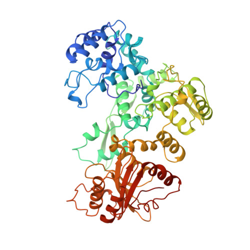
Structure of the multimodular endonuclease FokI bound to DNA.

Wah, D.A.Hirsch, J.A.Dorner, L.F.Schildkraut, I.Aggarwal, A.K.

(1997) Nature 388: 97-100

  • DOI: https://doi.org/10.1038/40446
  • Primary Citation Related Structures: 
    1FOK

  • PubMed Abstract: 

    FokI is a member of an unusual class of bipartite restriction enzymes that recognize a specific DNA sequence and cleave DNA nonspecifically a short distance away from that sequence. Because of its unusual bipartite nature, FokI has been used to create artificial enzymes with new specificities. We have determined the crystal structure at 2.8A resolution of the complete FokI enzyme bound to DNA. As anticipated, the enzyme contains amino- and carboxy-terminal domains corresponding to the DNA-recognition and cleavage functions, respectively. The recognition domain is made of three smaller subdomains (D1, D2 and D3) which are evolutionarily related to the helix-turn-helix-containing DNA-binding domain of the catabolite gene activator protein CAP. The CAP core has been extensively embellished in the first two subdomains, whereas in the third subdomain it has been co-opted for protein-protein interactions. Surprisingly, the cleavage domain contains only a single catalytic centre, raising the question of how monomeric FokI manages to cleave both DNA strands. Unexpectedly, the cleavage domain is sequestered in a 'piggyback' fashion by the recognition domain. The structure suggests a new mechanism for nuclease activation and provides a framework for the design of chimaeric enzymes with altered specificities.


  • Organizational Affiliation
    • Department of Biochemistry and Molecular Biophysics, Columbia University, New York 10032, USA.

Macromolecule Content 

  • Total Structure Weight: 77.79 kDa 
  • Atom Count: 5,527 
  • Modeled Residue Count: 608 
  • Deposited Residue Count: 616 
  • Unique protein chains: 1
  • Unique nucleic acid chains: 2

Macromolecules


Find similar proteins by:|  3D Structure
Entity ID: 3
MoleculeChains  Sequence LengthOrganismDetailsImage
PROTEIN (FOKI RESTRICTION ENDONUCLEASE)C [auth A]576Planomicrobium okeanokoitesMutation(s): 0 
EC: 3.1.21.4
UniProt
Find proteins for P14870 (Planomicrobium okeanokoites)
Explore P14870 
Go to UniProtKB:  P14870
Entity Groups
Sequence Clusters30% Identity50% Identity70% Identity90% Identity95% Identity100% Identity
UniProt GroupP14870
Sequence Annotations
Expand
Reference Sequence
Find similar nucleic acids by:  Sequence
Entity ID: 1
MoleculeChains LengthOrganismImage
DNA (5'-D(*TP*CP*GP*GP*AP*TP*GP*AP*TP*AP*AP*CP*GP*CP*TP*AP*G P*TP*CP*A)-3')A [auth B]20Planomicrobium okeanokoites
Sequence Annotations
Expand
Reference Sequence
Find similar nucleic acids by:  Sequence
Entity ID: 2
MoleculeChains LengthOrganismImage
DNA (5'-D(*AP*TP*GP*AP*CP*TP*AP*GP*CP*GP*TP*TP*AP*TP*CP*AP*T P*CP*CP*G)-3')B [auth C]20N/A
Sequence Annotations
Expand
Reference Sequence

Experimental Data & Validation

Experimental Data

  • Method: X-RAY DIFFRACTION
  • Resolution: 2.80 Å
  • R-Value Free:  0.296 (Depositor) 
  • R-Value Work:  0.214 (Depositor) 
  • R-Value Observed: 0.214 (Depositor) 
Space Group: P 1 21 1
Unit Cell:
Length ( Å )Angle ( ˚ )
a = 65.59α = 90
b = 119.34β = 101.42
c = 71.52γ = 90
Software Package:
Software NamePurpose
PHASESphasing
CCP4model building
SOLOMONphasing
X-PLORmodel building
X-PLORrefinement
DENZOdata reduction
SCALEPACKdata scaling
CCP4phasing
X-PLORphasing

Structure Validation

View Full Validation Report


Entry History 

Deposition Data

Revision History  (Full details and data files)

  • Version 1.0: 1997-12-03
    Type: Initial release
  • Version 1.1: 2008-05-22
    Changes: Version format compliance
  • Version 1.2: 2011-07-13
    Changes: Version format compliance
  • Version 1.3: 2021-06-30
    Changes: Data collection
  • Version 1.4: 2024-02-07
    Changes: Data collection, Database references