1FCA | pdb_00001fca

STRUCTURE OF THE FERREDOXIN FROM CLOSTRIDIUM ACIDURICI: MODEL AT 1.8 ANGSTROMS RESOLUTION


Experimental Data Snapshot

  • Method: X-RAY DIFFRACTION
  • Resolution: 1.80 Å
  • R-Value Work: 
    0.143 (Depositor) 
  • R-Value Observed: 
    0.143 (Depositor) 

wwPDB Validation 3D Report Full Report

Validation slider image for 1FCA

This is version 1.5 of the entry. See complete history

Literature

Structure of the ferredoxin from Clostridium acidurici: model at 1.8 A resolution.

TranQui, D.Jesior, J.C.

(1995) Acta Crystallogr D Biol Crystallogr 51: 155-159

  • DOI: https://doi.org/10.1107/S0907444994010735
  • Primary Citation Related Structures: 
    1FCA

  • PubMed Abstract: 

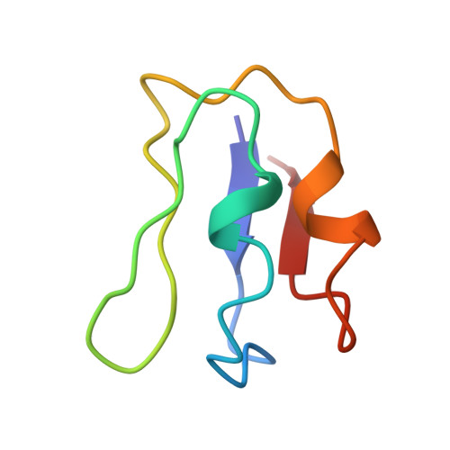
    Ferredoxins (Fd) are electron-carrier proteins, the active sites of which are organized around clusters made of iron and inorganic sulfur. The Fd from Clostridium acidurici is 55 amino acids long and contains two [4Fe-4S] clusters. Crystals have been obtained in the space group P4(3)2(1)2, a = b = 34.441 (5), c = 74.778 (9) A. The structure was solved by molecular replacement using the Fd from Peptostreptcoccus asaccharolyticus as a search model, these two ferredoxins having 37 residues in common. Refinement using molecular-dynamics techniques was then initiated. Successive rounds of model building and refinement gave a structure that includes 45 water molecules with R = 15%. At this stage, the electron-density map clearly revealed discrepancies in the position of two amino acids in the published primary sequence. Refinement based on these modifications led to R = 14.3% for 3921 reflections up to 1.8 A, resolution. The geometry of the two clusters has been found to be in good agreement with that previously obtained at a lower resolution. Interactions of polypeptide chain with the [4Fe-4S] clusters, the cluster geometry as well as the hydrogen bonds involving S, Sgamma, N and water molecules are reported.


  • Organizational Affiliation
    • Laboratoire de Cristallographie, CNRS, Grenoble, France.

Macromolecule Content 

  • Total Structure Weight: 6.2 kDa 
  • Atom Count: 438 
  • Modeled Residue Count: 55 
  • Deposited Residue Count: 55 
  • Unique protein chains: 1

Macromolecules

Find similar proteins by:|  3D Structure
Entity ID: 1
MoleculeChains  Sequence LengthOrganismDetailsImage
FERREDOXIN55Gottschalkia aciduriciMutation(s): 0 
UniProt
Find proteins for P00198 (Gottschalkia acidurici (strain ATCC 7906 / DSM 604 / BCRC 14475 / CIP 104303 / KCTC 5404 / NCIMB 10678 / 9a))
Explore P00198 
Go to UniProtKB:  P00198
Entity Groups
Sequence Clusters30% Identity50% Identity70% Identity90% Identity95% Identity100% Identity
UniProt GroupP00198
Sequence Annotations
Expand
Reference Sequence

Small Molecules

Ligands 1 Unique
IDChains Name / Formula / InChI Key2D Diagram3D Interactions
SF4

Query on SF4


Download:Ideal Coordinates CCD File
B [auth A],
C [auth A]
IRON/SULFUR CLUSTER
Fe4 S4
LJBDFODJNLIPKO-UHFFFAOYSA-N

Experimental Data & Validation

Experimental Data

  • Method: X-RAY DIFFRACTION
  • Resolution: 1.80 Å
  • R-Value Work:  0.143 (Depositor) 
  • R-Value Observed: 0.143 (Depositor) 
Space Group: P 43 21 2
Unit Cell:
Length ( Å )Angle ( ˚ )
a = 34.441α = 90
b = 34.441β = 90
c = 74.778γ = 90
Software Package:
Software NamePurpose
X-PLORmodel building
X-PLORrefinement
X-PLORphasing

Structure Validation

View Full Validation Report


Entry History 

Deposition Data

Revision History  (Full details and data files)

  • Version 1.0: 1995-07-10
    Type: Initial release
  • Version 1.1: 2008-03-03
    Changes: Version format compliance
  • Version 1.2: 2011-07-13
    Changes: Version format compliance
  • Version 1.3: 2011-11-16
    Changes: Atomic model
  • Version 1.4: 2017-11-29
    Changes: Derived calculations, Other
  • Version 1.5: 2024-02-07
    Changes: Data collection, Database references, Derived calculations