


Combined Rietveld and Stereochemical Restraint Refinement of a Protein Crystal Structure
Von Dreele, R.B.(1999) J Appl Crystallogr 32: 1084-1089
Experimental Data Snapshot
wwPDB Validation 3D Report Full Report

(1999) J Appl Crystallogr 32: 1084-1089
Entity ID: 1 | |||||
|---|---|---|---|---|---|
| Molecule | Chains | Sequence Length | Organism | Details | Image |
| MYOGLOBIN (MET) | 153 | Physeter macrocephalus | Mutation(s): 0 EC: 1.11.1 (UniProt), 1.7 (UniProt) | 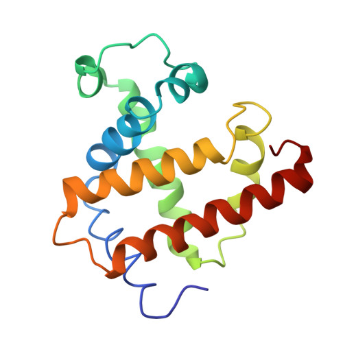 | |
UniProt | |||||
Entity Groups | |||||
| Sequence Clusters | 30% Identity50% Identity70% Identity90% Identity95% Identity100% Identity | ||||
| UniProt Group | P02185 | ||||
Sequence AnnotationsExpand | |||||
Reference Sequence | |||||
| Ligands 1 Unique | |||||
|---|---|---|---|---|---|
| ID | Chains | Name / Formula / InChI Key | 2D Diagram | 3D Interactions | |
| HEM Download:Ideal Coordinates CCD File | B [auth A] | PROTOPORPHYRIN IX CONTAINING FE C34 H32 Fe N4 O4 KABFMIBPWCXCRK-RGGAHWMASA-L |  | ||
| Length ( Å ) | Angle ( ˚ ) |
|---|---|
| a = 64.4113 | α = 90 |
| b = 30.1946 | β = 105.834 |
| c = 34.9476 | γ = 90 |
| Software Name | Purpose |
|---|---|
| GSAS | refinement |
| PROCESS | data scaling |


