1F5L

UROKINASE PLASMINOGEN ACTIVATOR B-CHAIN-AMILORIDE COMPLEX


Experimental Data Snapshot

  • Method: X-RAY DIFFRACTION
  • Resolution: 2.10 Å
  • R-Value Free: 0.250 
  • R-Value Work: 0.202 

wwPDB Validation   3D Report Full Report


This is version 1.3 of the entry. See complete history


Literature

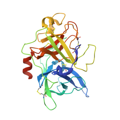
Crystals of the urokinase type plasminogen activator variant beta(c)-uPAin complex with small molecule inhibitors open the way towards structure-based drug design.

Zeslawska, E.Schweinitz, A.Karcher, A.Sondermann, P.Sperl, S.Sturzebecher, J.Jacob, U.

(2000) J Mol Biol 301: 465-475

  • DOI: https://doi.org/10.1006/jmbi.2000.3966
  • Primary Citation of Related Structures:  
    1F5K, 1F5L, 1F92

  • PubMed Abstract: 

    Urokinase is a serine protease involved in cancer growth and metastasis. Here we present the first urokinase crystal structure in complex with reversible inhibitors at 2.1 and 2.6 A resolution. These inhibitor complex structures have been obtained from crystals of engineered urokinase type plasminogen activator designed to obtain a crystal form open for inhibitor soaking. The mutant C122S loses its flexible A-chain upon activation cleavage and crystallizes in the presence of benzamidine, which was later displaced by the desired inhibitor. This new soakable crystal form turned out to be of great value in the process of structure-based drug design. The evaluated binding mode of amiloride, and UKI-1D revealed a new subsite of the primary specificity pocket of urokinase that will be employed in the future ligand optimisation process.


  • Organizational Affiliation

    Max-Planck-Institut für Biochemie, Abteilung Strukturforschung, Martinsried, Am Klopferspitz 18a, D-82152, Germany.


Macromolecules
Find similar proteins by:  (by identity cutoff)  |  3D Structure
Entity ID: 1
MoleculeChains Sequence LengthOrganismDetailsImage
UROKINASE-TYPE PLASMINOGEN ACTIVATOR253Homo sapiensMutation(s): 1 
EC: 3.4.21.73
UniProt & NIH Common Fund Data Resources
Find proteins for P00749 (Homo sapiens)
Explore P00749 
Go to UniProtKB:  P00749
PHAROS:  P00749
GTEx:  ENSG00000122861 
Entity Groups  
Sequence Clusters30% Identity50% Identity70% Identity90% Identity95% Identity100% Identity
UniProt GroupP00749
Sequence Annotations
Expand
  • Reference Sequence
Small Molecules
Ligands 2 Unique
IDChains Name / Formula / InChI Key2D Diagram3D Interactions
AMR
Query on AMR

Download Ideal Coordinates CCD File 
D [auth A]3,5-DIAMINO-N-(AMINOIMINOMETHYL)-6-CHLOROPYRAZINECARBOXAMIDE
C6 H8 Cl N7 O
XSDQTOBWRPYKKA-UHFFFAOYSA-N
SO4
Query on SO4

Download Ideal Coordinates CCD File 
B [auth A],
C [auth A]
SULFATE ION
O4 S
QAOWNCQODCNURD-UHFFFAOYSA-L
Binding Affinity Annotations 
IDSourceBinding Affinity
AMR BindingDB:  1F5L Ki: min: 2433, max: 7000 (nM) from 4 assay(s)
IC50: min: 2400, max: 1.07e+5 (nM) from 4 assay(s)
Binding MOAD:  1F5L Ki: 5300 (nM) from 1 assay(s)
PDBBind:  1F5L Ki: 5300 (nM) from 1 assay(s)
Experimental Data & Validation

Experimental Data

  • Method: X-RAY DIFFRACTION
  • Resolution: 2.10 Å
  • R-Value Free: 0.250 
  • R-Value Work: 0.202 
  • Space Group: P 21 21 21
Unit Cell:
Length ( Å )Angle ( ˚ )
a = 52.8α = 90
b = 54.6β = 90
c = 82.92γ = 90
Software Package:
Software NamePurpose
MOSFLMdata reduction
SCALAdata scaling
AMoREphasing
CNSrefinement
CCP4data scaling

Structure Validation

View Full Validation Report


Entry History 

Deposition Data

Revision History  (Full details and data files)

  • Version 1.0: 2001-06-15
    Type: Initial release
  • Version 1.1: 2008-04-27
    Changes: Version format compliance
  • Version 1.2: 2011-07-13
    Changes: Version format compliance
  • Version 1.3: 2021-11-03
    Changes: Database references, Derived calculations