1F4K

CRYSTAL STRUCTURE OF THE REPLICATION TERMINATOR PROTEIN/B-SITE DNA COMPLEX


Experimental Data Snapshot

  • Method: X-RAY DIFFRACTION
  • Resolution: 2.50 Å
  • R-Value Free: 0.276 
  • R-Value Work: 0.228 
  • R-Value Observed: 0.271 

wwPDB Validation   3D Report Full Report


This is version 1.4 of the entry. See complete history


Literature

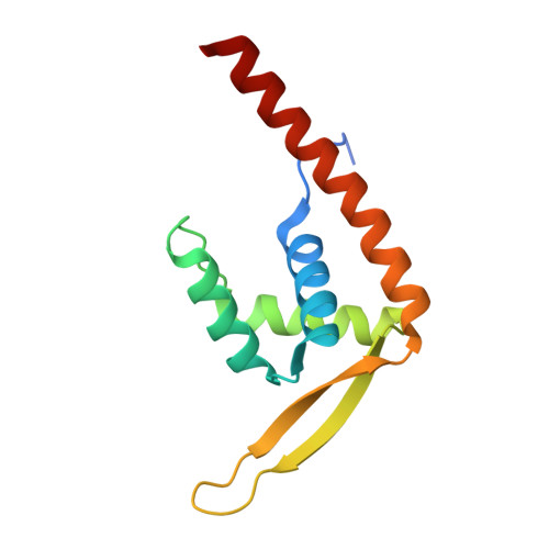
Structure of the RTP-DNA complex and the mechanism of polar replication fork arrest

Wilce, J.A.Vivian, J.P.Hastings, A.F.Otting, G.Folmer, R.H.Duggin, I.G.Wake, R.G.Wilce, M.C.

(2001) Nat Struct Biol 8: 206-210

  • DOI: https://doi.org/10.1038/84934
  • Primary Citation of Related Structures:  
    1F4K

  • PubMed Abstract: 

    The coordinated termination of DNA replication is an important step in the life cycle of bacteria with circular chromosomes, but has only been defined at a molecular level in two systems to date. Here we report the structure of an engineered replication terminator protein (RTP) of Bacillus subtilis in complex with a 21 base pair DNA by X-ray crystallography at 2.5 A resolution. We also use NMR spectroscopic titration techniques. This work reveals a novel DNA interaction involving a dimeric 'winged helix' domain protein that differs from predictions. While the two recognition helices of RTP are in close contact with the B-form DNA major grooves, the 'wings' and N-termini of RTP do not form intimate contacts with the DNA. This structure provides insight into the molecular basis of polar replication fork arrest based on a model of cooperative binding and differential binding affinities of RTP to the two adjacent binding sites in the complete terminator.


  • Organizational Affiliation

    Department of Chemistry/Biochemistry University of Western Australia and the Western Australian Institute for Medical Research, Nedlands, Western Australia 6907 Australia.


Macromolecules

Find similar proteins by:  (by identity cutoff)  |  3D Structure
Entity ID: 3
MoleculeChains Sequence LengthOrganismDetailsImage
REPLICATION TERMINATION PROTEINC [auth A],
D [auth B]
122Bacillus subtilisMutation(s): 1 
UniProt
Find proteins for P0CI76 (Bacillus subtilis (strain 168))
Explore P0CI76 
Go to UniProtKB:  P0CI76
Entity Groups  
Sequence Clusters30% Identity50% Identity70% Identity90% Identity95% Identity100% Identity
UniProt GroupP0CI76
Sequence Annotations
Expand
  • Reference Sequence

Find similar nucleic acids by:  Sequence   |   3D Structure  

Entity ID: 1
MoleculeChains LengthOrganismImage
5'-D(*CP*TP*AP*TP*GP*AP*AP*CP*AP*TP*AP*AP*TP*GP*TP*TP*CP*AP*TP*AP*G)-3'A [auth D]21N/A
Sequence Annotations
Expand
  • Reference Sequence

Find similar nucleic acids by:  Sequence   |   3D Structure  

Entity ID: 2
MoleculeChains LengthOrganismImage
5'-D(*CP*TP*AP*TP*GP*AP*AP*CP*AP*TP*TP*AP*TP*GP*TP*TP*CP*AP*TP*AP*G)-3'B [auth E]21N/A
Sequence Annotations
Expand
  • Reference Sequence
Experimental Data & Validation

Experimental Data

  • Method: X-RAY DIFFRACTION
  • Resolution: 2.50 Å
  • R-Value Free: 0.276 
  • R-Value Work: 0.228 
  • R-Value Observed: 0.271 
  • Space Group: P 32 2 1
Unit Cell:
Length ( Å )Angle ( ˚ )
a = 44.78α = 90
b = 44.78β = 90
c = 395.58γ = 120
Software Package:
Software NamePurpose
DENZOdata reduction
SCALEPACKdata scaling
AMoREphasing
CNSrefinement

Structure Validation

View Full Validation Report


Entry History 

Deposition Data

Revision History  (Full details and data files)

  • Version 1.0: 2001-06-08
    Type: Initial release
  • Version 1.1: 2008-04-27
    Changes: Version format compliance
  • Version 1.2: 2011-07-13
    Changes: Version format compliance
  • Version 1.3: 2021-11-03
    Changes: Database references
  • Version 1.4: 2024-02-07
    Changes: Data collection