1F3H | pdb_00001f3h

X-RAY CRYSTAL STRUCTURE OF THE HUMAN ANTI-APOPTOTIC PROTEIN SURVIVIN


Experimental Data Snapshot

  • Method: X-RAY DIFFRACTION
  • Resolution: 2.58 Å
  • R-Value Free: 
    0.287 (Depositor) 
  • R-Value Work: 
    0.228 (Depositor) 
  • R-Value Observed: 
    0.228 (Depositor) 

wwPDB Validation 3D Report Full Report

Validation slider image for 1F3H

This is version 1.4 of the entry. See complete history

Literature

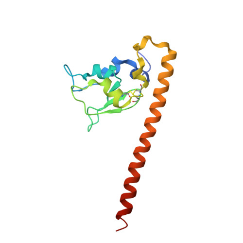
Structure of the human anti-apoptotic protein survivin reveals a dimeric arrangement.

Verdecia, M.A.Huang, H.Dutil, E.Kaiser, D.A.Hunter, T.Noel, J.P.

(2000) Nat Struct Biol 7: 602-608

  • DOI: https://doi.org/10.1038/76838
  • Primary Citation Related Structures: 
    1F3H

  • PubMed Abstract: 

    Survivin is a 16.5 kDa protein that is expressed during the G2/M phase of the cell cycle and is hypothesized to inhibit a default apoptotic cascade initiated in mitosis. This inhibitory function is coupled to survivin's localization to the mitotic spindle. To begin to address the structural basis of survivin's function, we report the X-ray crystal structure of a recombinant form of full length survivin to 2.58 A resolution. Survivin consists of two defined domains including an N-terminal Zn2+-binding BIR domain linked to a 65 A amphipathic C-terminal alpha-helix. The crystal structure reveals an extensive dimerization interface along a hydrophobic surface on the BIR domain of each survivin monomer. A basic patch acting as a sulfate/phosphate-binding module, an acidic cluster projecting off the BIR domain, and a solvent-accessible hydrophobic surface residing on the C-terminal amphipathic helix, are suggestive of functional protein-protein interaction surfaces.


  • Organizational Affiliation
    • Structural Biology Laboratory, The Salk Institute for Biological Studies, La Jolla, California 92037, USA.

Macromolecule Content 

  • Total Structure Weight: 33.28 kDa 
  • Atom Count: 2,320 
  • Modeled Residue Count: 271 
  • Deposited Residue Count: 284 
  • Unique protein chains: 1

Macromolecules

Find similar proteins by:|  3D Structure
Entity ID: 1
MoleculeChains  Sequence LengthOrganismDetailsImage
SURVIVIN
A, B
142Homo sapiensMutation(s): 1 
UniProt & NIH Common Fund Data Resources
Find proteins for O15392 (Homo sapiens)
Explore O15392 
Go to UniProtKB:  O15392
PHAROS:  O15392
GTEx:  ENSG00000089685 
Entity Groups
Sequence Clusters30% Identity50% Identity70% Identity90% Identity95% Identity100% Identity
UniProt GroupO15392
Sequence Annotations
Expand
Reference Sequence

Experimental Data & Validation

Experimental Data

  • Method: X-RAY DIFFRACTION
  • Resolution: 2.58 Å
  • R-Value Free:  0.287 (Depositor) 
  • R-Value Work:  0.228 (Depositor) 
  • R-Value Observed: 0.228 (Depositor) 
Space Group: C 1 2 1
Unit Cell:
Length ( Å )Angle ( ˚ )
a = 114.04α = 90
b = 71.45β = 133.37
c = 86.63γ = 90
Software Package:
Software NamePurpose
DENZOdata reduction
SCALEPACKdata scaling
SHARPphasing
CNSrefinement

Structure Validation

View Full Validation Report


Entry History 

Deposition Data

Revision History  (Full details and data files)

  • Version 1.0: 2000-12-06
    Type: Initial release
  • Version 1.1: 2008-04-27
    Changes: Version format compliance
  • Version 1.2: 2011-07-13
    Changes: Version format compliance
  • Version 1.3: 2021-11-03
    Changes: Database references, Derived calculations
  • Version 1.4: 2024-11-06
    Changes: Data collection, Structure summary