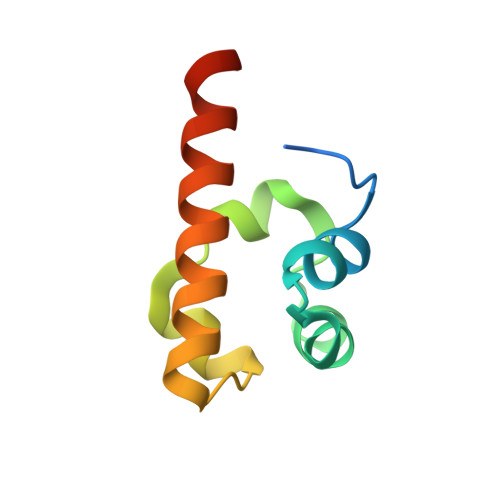
Monomeric structure of the human EphB2 sterile alpha motif domain.
Thanos, C.D., Faham, S., Goodwill, K.E., Cascio, D., Phillips, M., Bowie, J.U.(1999) J Biological Chem 274: 37301-37306
- PubMed: 10601296 Search on PubMed
- DOI: https://doi.org/10.1074/jbc.274.52.37301
- Primary Citation Related Structures:
1F0M - PubMed Abstract:
The sterile alpha motif (SAM) domain is a protein module found in many diverse signaling proteins. SAM domains in some systems have been shown to self-associate. Previous crystal structures of an EphA4-SAM domain dimer (Stapleton, D., Balan, I., Pawson, T., and Sicheri, F. (1999) Nat. Struct. Biol. 6, 44-49) and a possible EphB2-SAM oligomer (Thanos, C. D., Goodwill, K. E., and Bowie, J. U. (1999) Science 283, 833-836) both revealed large interfaces comprising an exchange of N-terminal peptide arms. Within the arm, a conserved hydrophobic residue (Tyr-8 in the EphB2-SAM structure or Phe-910 in the EphA4-SAM structure) is anchored into a hydrophobic cleft on a neighboring molecule. Here we have solved a new crystal form of the human EphB2-SAM domain that has the same overall SAM domain fold yet has no substantial intermolecular contacts. In the new structure, the N-terminal peptide arm of the EphB2-SAM domain protrudes out from the core of the molecule, leaving both the arm (including Tyr-8) and the hydrophobic cleft solvent-exposed. To verify that Tyr-8 is solvent-exposed in solution, we made a Tyr-8 to Ala-8 mutation and found that the EphB2-SAM domain structure and stability were only slightly altered. These results suggest that Tyr-8 is not part of the hydrophobic core of the EphB2-SAM domain and is conserved for functional reasons. Cystallographic evidence suggests a possible role for the N-terminal arm in oligomerization. In the absence of a direct demonstration of biological relevance, however, the functional role of the N-terminal arm remains an open question.
- UCLA-DOE Laboratory of Structural Biology, Department of Chemistry and Biochemistry, UCLA, Los Angeles, California 90095, USA.
Organizational Affiliation: