1EXU

CRYSTAL STRUCTURE OF THE HUMAN MHC-RELATED FC RECEPTOR


Experimental Data Snapshot

  • Method: X-RAY DIFFRACTION
  • Resolution: 2.70 Å
  • R-Value Free: 0.247 
  • R-Value Work: 0.197 

wwPDB Validation   3D Report Full Report


This is version 2.0 of the entry. See complete history


Literature

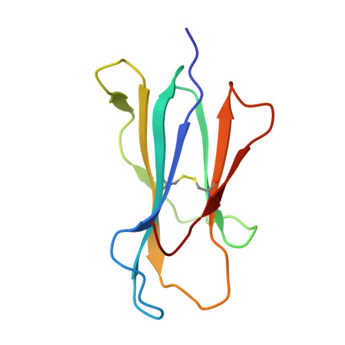
Crystal structure and immunoglobulin G binding properties of the human major histocompatibility complex-related Fc receptor(,).

West Jr., A.P.Bjorkman, P.J.

(2000) Biochemistry 39: 9698-9708

  • DOI: https://doi.org/10.1021/bi000749m
  • Primary Citation of Related Structures:  
    1EXU

  • PubMed Abstract: 

    The neonatal Fc receptor (FcRn) performs two distinct but related functions: transport of maternal immunoglobulin G (IgG) to pre- or neonatal mammals, thus providing passive immunity, and protection of IgG from normal serum protein catabolism. FcRn is related to class I MHC proteins but lacks a functional peptide binding groove. The crystal structure of human FcRn has been determined at 2.7 A resolution and compared to the previously described structure of rat FcRn [Burmeister et al. (1994) Nature 372, 336-343] and to the structures of MHC and MHC-related proteins. Human FcRn is structurally similar to the rat receptor but does not form receptor dimers in the crystals as observed in crystals of rat FcRn. The interaction between human FcRn and IgG was characterized by determining the binding stoichiometry using equilibrium gel filtration and by deriving binding affinities for the different human IgG subclasses using a surface plasmon resonance assay. Like rat and mouse FcRn, human FcRn interacts with IgG with a 2:1 receptor:ligand stoichiometry. The binding of human FcRn to the four human IgG subclasses shows subclass and allotype variations but no clear subclass affinity differences that correlate with serum half-lives. The structure of human FcRn and studies of its ligand binding are relevant to current efforts to use FcRn-mediated regulation of IgG half-life in serum to increase the lifetimes of antibody-based therapeutics.


  • Organizational Affiliation

    Division of Biology 156-29 and Howard Hughes Medical Institute, California Institute of Technology, Pasadena, California 91125, USA.


Macromolecules
Find similar proteins by:  (by identity cutoff)  |  3D Structure
Entity ID: 1
MoleculeChains Sequence LengthOrganismDetailsImage
IGG RECEPTOR FCRN LARGE SUBUNIT P51267Homo sapiensMutation(s): 0 
UniProt & NIH Common Fund Data Resources
Find proteins for P55899 (Homo sapiens)
Explore P55899 
Go to UniProtKB:  P55899
PHAROS:  P55899
GTEx:  ENSG00000104870 
Entity Groups  
Sequence Clusters30% Identity50% Identity70% Identity90% Identity95% Identity100% Identity
UniProt GroupP55899
Sequence Annotations
Expand
  • Reference Sequence
Find similar proteins by:  (by identity cutoff)  |  3D Structure
Entity ID: 2
MoleculeChains Sequence LengthOrganismDetailsImage
BETA-2-MICROGLOBULIN99Homo sapiensMutation(s): 0 
UniProt & NIH Common Fund Data Resources
Find proteins for P61769 (Homo sapiens)
Explore P61769 
Go to UniProtKB:  P61769
PHAROS:  P61769
GTEx:  ENSG00000166710 
Entity Groups  
Sequence Clusters30% Identity50% Identity70% Identity90% Identity95% Identity100% Identity
UniProt GroupP61769
Sequence Annotations
Expand
  • Reference Sequence
Small Molecules
Ligands 1 Unique
IDChains Name / Formula / InChI Key2D Diagram3D Interactions
BME
Query on BME

Download Ideal Coordinates CCD File 
C [auth A]BETA-MERCAPTOETHANOL
C2 H6 O S
DGVVWUTYPXICAM-UHFFFAOYSA-N
Experimental Data & Validation

Experimental Data

  • Method: X-RAY DIFFRACTION
  • Resolution: 2.70 Å
  • R-Value Free: 0.247 
  • R-Value Work: 0.197 
  • Space Group: P 43 21 2
Unit Cell:
Length ( Å )Angle ( ˚ )
a = 66.387α = 90
b = 66.387β = 90
c = 221.813γ = 90
Software Package:
Software NamePurpose
DENZOdata reduction
SCALEPACKdata scaling
AMoREphasing
X-PLORrefinement

Structure Validation

View Full Validation Report


Entry History 

Deposition Data

Revision History  (Full details and data files)

  • Version 1.0: 2000-08-10
    Type: Initial release
  • Version 1.1: 2008-04-27
    Changes: Version format compliance
  • Version 1.2: 2011-07-13
    Changes: Version format compliance
  • Version 2.0: 2023-08-09
    Changes: Atomic model, Data collection, Database references, Derived calculations, Refinement description