1EVR

The structure of the resorcinol/insulin R6 hexamer


Experimental Data Snapshot

  • Method: X-RAY DIFFRACTION
  • Resolution: 1.90 Å
  • R-Value Free: 0.218 
  • R-Value Work: 0.179 
  • R-Value Observed: 0.179 

wwPDB Validation   3D Report Full Report


This is version 1.3 of the entry. See complete history


Literature

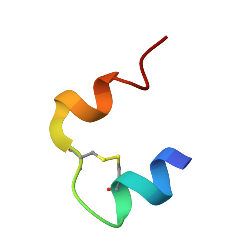
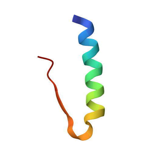
R6 hexameric insulin complexed with m-cresol or resorcinol.

Smith, G.D.Ciszak, E.Magrum, L.A.Pangborn, W.A.Blessing, R.H.

(2000) Acta Crystallogr D Biol Crystallogr 56: 1541-1548

  • DOI: https://doi.org/10.1107/s0907444900012749
  • Primary Citation of Related Structures:  
    1EV3, 1EV6, 1EVR

  • PubMed Abstract: 
  • The structures of three R(6) human insulin hexamers have been determined. Crystals of monoclinic m-cresol-insulin, monoclinic resorcinol-insulin and rhombohedral m-cresol-insulin diffracted to 1. 9, 1.9 and 1.78 A, respectively, and have been refined to residuals of 0 ...

    The structures of three R(6) human insulin hexamers have been determined. Crystals of monoclinic m-cresol-insulin, monoclinic resorcinol-insulin and rhombohedral m-cresol-insulin diffracted to 1. 9, 1.9 and 1.78 A, respectively, and have been refined to residuals of 0.195, 0.179 and 0.200, respectively. In all three structures, a phenolic derivative is found to occupy the phenolic binding site, where it forms hydrogen bonds to the carbonyl O atom of CysA6 and the N atom of CysA11. Two additional phenolic derivative binding sites were identified within or between hexamers. The structures of all three hexamers are nearly identical, although a large displacement of the N-terminus of one B chain in both monoclinic structures results from coordination to a sodium ion which is located between symmetry-related hexamers. Other minor differences in structure arise from differences in packing in the monoclinic cell compared with the rhombohedral cell. Based upon the differences in conformation of the GluB13 side chains in T(6), T(3)R(f)(3) and R(6) hexamers, the deprotonation of these side chains appears to be associated with the T-->R conformational transition.


    Related Citations: 
    • Structure of a Rhombohedral R6 Insuliin/Phenol Complex
      Smith, G.D., Dodson, G.G.
      (1992) Proteins 14: 401
    • Phenol Stabilizes more Helix in a New Symmetrical Zinc Insulin Hexamer
      Derewenda, U., Derewenda, Z., Dodson, E.J., Dodson, G.G., Reynolds, C.D., Smith, G.D., Sparks, C., Swenson, D.
      (1989) Nature 338: 594

    Organizational Affiliation

    Hauptman-Woodward Medical Research Institute, 73 High Street, Buffalo, NY 14203, USA. smith@hwi.buffalo.edu


Macromolecules

Find similar proteins by:  Sequence   |   3D Structure  

Entity ID: 1
MoleculeChainsSequence LengthOrganismDetailsImage
INSULIN
A, C, E, G, I, K
A, C, E, G, I, K
21N/AMutation(s): 0 
UniProt & NIH Common Fund Data Resources
Find proteins for P01308 (Homo sapiens)
Explore P01308 
Go to UniProtKB:  P01308
PHAROS:  P01308
Entity Groups  
Sequence Clusters30% Identity50% Identity70% Identity90% Identity95% Identity100% Identity
UniProt GroupP01308
Protein Feature View
Expand
  • Reference Sequence
Find similar proteins by:  (by identity cutoff)  |  3D Structure
Entity ID: 2
MoleculeChainsSequence LengthOrganismDetailsImage
INSULIN
B, D, F, H, J, L
B, D, F, H, J, L
30N/AMutation(s): 0 
UniProt & NIH Common Fund Data Resources
Find proteins for P01308 (Homo sapiens)
Explore P01308 
Go to UniProtKB:  P01308
PHAROS:  P01308
Entity Groups  
Sequence Clusters30% Identity50% Identity70% Identity90% Identity95% Identity100% Identity
UniProt GroupP01308
Protein Feature View
Expand
  • Reference Sequence
Small Molecules
Ligands 4 Unique
IDChainsName / Formula / InChI Key2D Diagram3D Interactions
RCO
Query on RCO

Download Ideal Coordinates CCD File 
M [auth A],
N [auth A],
P [auth B],
Q [auth C],
T [auth E],
M [auth A],
N [auth A],
P [auth B],
Q [auth C],
T [auth E],
U [auth G],
V [auth I],
Y [auth K]
RESORCINOL
C6 H6 O2
GHMLBKRAJCXXBS-UHFFFAOYSA-N
 Ligand Interaction
ZN
Query on ZN

Download Ideal Coordinates CCD File 
O [auth B],
R [auth D]
ZINC ION
Zn
PTFCDOFLOPIGGS-UHFFFAOYSA-N
 Ligand Interaction
CL
Query on CL

Download Ideal Coordinates CCD File 
S [auth D],
W [auth J]
CHLORIDE ION
Cl
VEXZGXHMUGYJMC-UHFFFAOYSA-M
 Ligand Interaction
NA
Query on NA

Download Ideal Coordinates CCD File 
X [auth J]SODIUM ION
Na
FKNQFGJONOIPTF-UHFFFAOYSA-N
 Ligand Interaction
Experimental Data & Validation

Experimental Data

  • Method: X-RAY DIFFRACTION
  • Resolution: 1.90 Å
  • R-Value Free: 0.218 
  • R-Value Work: 0.179 
  • R-Value Observed: 0.179 
  • Space Group: P 1 21 1
Unit Cell:
Length ( Å )Angle ( ˚ )
a = 61.35α = 90
b = 61.926β = 110.61
c = 47.805γ = 90
Software Package:
Software NamePurpose
CNSrefinement
CNSphasing

Structure Validation

View Full Validation Report


Entry History 

Deposition Data

Revision History  (Full details and data files)

  • Version 1.0: 2000-12-04
    Type: Initial release
  • Version 1.1: 2008-04-27
    Changes: Version format compliance
  • Version 1.2: 2011-07-13
    Changes: Version format compliance
  • Version 1.3: 2017-10-04
    Changes: Refinement description