1ETH | pdb_00001eth

TRIACYLGLYCEROL LIPASE/COLIPASE COMPLEX


Experimental Data Snapshot

  • Method: X-RAY DIFFRACTION
  • Resolution: 2.80 Å
  • R-Value Free: 
    0.290 (Depositor) 
  • R-Value Work: 
    0.210 (Depositor) 
  • R-Value Observed: 
    0.210 (Depositor) 

wwPDB Validation 3D Report Full Report

Validation slider image for 1ETH

This is version 2.1 of the entry. See complete history

Literature

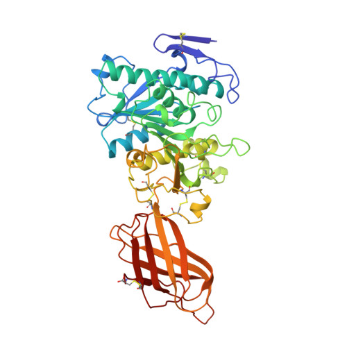
Lipase activation by nonionic detergents. The crystal structure of the porcine lipase-colipase-tetraethylene glycol monooctyl ether complex.

Hermoso, J.Pignol, D.Kerfelec, B.Crenon, I.Chapus, C.Fontecilla-Camps, J.C.

(1996) J Biological Chem 271: 18007-18016

  • DOI: https://doi.org/10.1074/jbc.271.30.18007
  • Primary Citation Related Structures: 
    1ETH

  • PubMed Abstract: 

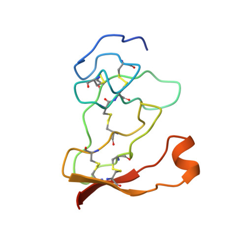
    The crystal structure of the ternary porcine lipase-colipase-tetra ethylene glycol monooctyl ether (TGME) complex has been determined at 2.8 A resolution. The crystals belong to the cubic space group F23 with a = 289.1 A and display a strong pseudo-symmetry corresponding to a P23 lattice. Unexpectedly, the crystalline two-domain lipase is found in its open configuration. This indicates that in the presence of colipase, pure micelles of the nonionic detergent TGME are able to activate the enzyme; a process that includes the movement of an N-terminal domain loop (the flap). The effects of TGME and colipase have been confirmed by chemical modification of the active site serine residue using diisopropyl p-nitrophenylphosphate (E600). In addition, the presence of a TGME molecule tightly bound to the active site pocket shows that TGME acts as a substrate analog, thus possibly explaining the inhibitory effect of this nonionic detergent on emulsified substrate hydrolysis at submicellar concentrations. A comparison of the lipase-colipase interactions between our porcine complex and the human-porcine complex (van Tilbeurgh, H., Egloff, M.-P., Martinez, C., Rugani, N., Verger, R., and Cambillau, C.(1993) Nature 362, 814-820) indicates that except for one salt bridge interaction, they are conserved. Analysis of the superimposed complexes shows a 5.4 degrees rotation on the relative position of the N-terminal domains excepting the flap that moves in a concerted fashion with the C-terminal domain. This flexibility may be important for the binding of the complex to the water-lipid interface.


  • Organizational Affiliation
    • Laboratoire de Cristallographie et de Cristallogénèse des Protéines, Institut de Biologie Structurale Jean-Pierre Ebel, CEA-CNRS, Grenoble, France.

Macromolecule Content 

  • Total Structure Weight: 123.74 kDa 
  • Atom Count: 8,911 
  • Modeled Residue Count: 1,070 
  • Deposited Residue Count: 1,086 
  • Unique protein chains: 2

Macromolecules

Find similar proteins by:|  3D Structure
Entity ID: 1
MoleculeChains  Sequence LengthOrganismDetailsImage
TRIACYLGLYCEROL ACYL-HYDROLASE
A, C
448Sus scrofaMutation(s): 0 
EC: 3.1.1.3
UniProt
Find proteins for P00591 (Sus scrofa)
Explore P00591 
Go to UniProtKB:  P00591
Entity Groups
Sequence Clusters30% Identity50% Identity70% Identity90% Identity95% Identity100% Identity
UniProt GroupP00591
Glycosylation
Glycosylation Sites: 1
Sequence Annotations
Expand
Reference Sequence
Find similar proteins by:|  3D Structure
Entity ID: 2
MoleculeChains  Sequence LengthOrganismDetailsImage
COLIPASE
B, D
95Sus scrofaMutation(s): 0 
Membrane Entity: Yes 
UniProt
Find proteins for P02703 (Sus scrofa)
Explore P02703 
Go to UniProtKB:  P02703
Entity Groups
Sequence Clusters30% Identity50% Identity70% Identity90% Identity95% Identity100% Identity
UniProt GroupP02703
Sequence Annotations
Expand
Reference Sequence

Oligosaccharides

Help  
Entity ID: 3
MoleculeChains Length2D Diagram GlycosylationD Interactions
beta-D-mannopyranose-(1-3)-[beta-D-mannopyranose-(1-6)]beta-D-mannopyranose-(1-4)-2-acetamido-2-deoxy-beta-D-glucopyranose-(1-4)-2-acetamido-2-deoxy-beta-D-glucopyranose
E, F
5N-Glycosylation
Glycosylation Resources
GlyTouCan: G89225LT
GlyCosmos: G89225LT
GlyGen: G89225LT

Experimental Data & Validation

Experimental Data

  • Method: X-RAY DIFFRACTION
  • Resolution: 2.80 Å
  • R-Value Free:  0.290 (Depositor) 
  • R-Value Work:  0.210 (Depositor) 
  • R-Value Observed: 0.210 (Depositor) 
Space Group: F 2 3
Unit Cell:
Length ( Å )Angle ( ˚ )
a = 289.1α = 90
b = 289.1β = 90
c = 289.1γ = 90
Software Package:
Software NamePurpose
MOSFLMdata reduction
X-PLORmodel building
X-PLORrefinement
X-PLORphasing

Structure Validation

View Full Validation Report


Entry History 

Deposition Data

Revision History  (Full details and data files)

  • Version 1.0: 1996-12-07
    Type: Initial release
  • Version 1.1: 2008-03-27
    Changes: Version format compliance
  • Version 1.2: 2011-07-13
    Changes: Non-polymer description, Version format compliance
  • Version 1.3: 2018-03-21
    Changes: Data collection
  • Version 2.0: 2020-07-29
    Type: Remediation
    Reason: Carbohydrate remediation
    Changes: Advisory, Atomic model, Data collection, Database references, Derived calculations, Structure summary
  • Version 2.1: 2025-03-26
    Changes: Data collection, Database references, Structure summary