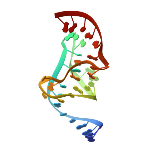
A water channel in the core of the vitamin B(12) RNA aptamer.
Sussman, D., Wilson, C.(2000) Structure 8: 719-727
- PubMed: 10903943 Search on PubMed
- DOI: https://doi.org/10.1016/s0969-2126(00)00159-3
- Primary Citation Related Structures:
1ET4 - PubMed Abstract:
The 3.0 A crystal structure of the vitamin B(12) RNA aptamer revealed an unusual tertiary structure that is rich in novel RNA structural motifs. Important details of the interactions that stabilize noncanonical base pairing and the role of solvent in the structure were not apparent owing to the limited resolution. The structure of the vitamin B(12) RNA aptamer in complex with its ligand has been determined at 2.3 A resolution by X-ray crystallography. The crystallographic asymmetric unit contains five independent copies of the aptamer-vitamin B(12) complex, making it possible to accurately define well-conserved features. The core of the aptamer contains an unusual water-filled channel that is buried between the three strands of an RNA triplex. Well-ordered water molecules positioned within this channel form bridging hydrogen bonds and stabilize planar base triples that otherwise lack significant direct base-base contacts. The water channel terminates at the interface between the RNA and the bound ligand, leaving a pair of water molecules appropriately positioned to hydrogen bond with the highly polarized cyanide nitrogen of vitamin B(12). Analysis of the general solvation patterns for each nucleotide suggests that water molecules are not precisely positioned, as observed in previous RNA duplex structures, but instead might adjust in response to the varying local environment. Unusual intermolecular base pairing contributes to the formation of three different dimerization contacts that drive formation of the crystal lattice. The structure demonstrates the important role of water molecules and noncanonical base pairing in driving the formation of RNA tertiary structure and facilitating specific interactions of RNAs with other molecules.
- Department of Biology, Center for the Molecular Biology of RNA, University of California at Santa Cruz, Santa Cruz, CA 95064, USA.
Organizational Affiliation: