TEM1 beta-lactamase structure solved by molecular replacement and refined structure of the S235A mutant.
Fonze, E., Charlier, P., To'th, Y., Vermeire, M., Raquet, X., Dubus, A., Frere, J.M.(1995) Acta Crystallogr D Biol Crystallogr 51: 682-694
- PubMed: 15299797 Search on PubMed
- DOI: https://doi.org/10.1107/S0907444994014496
- Primary Citation Related Structures:
1ESU, 1XPB - PubMed Abstract:
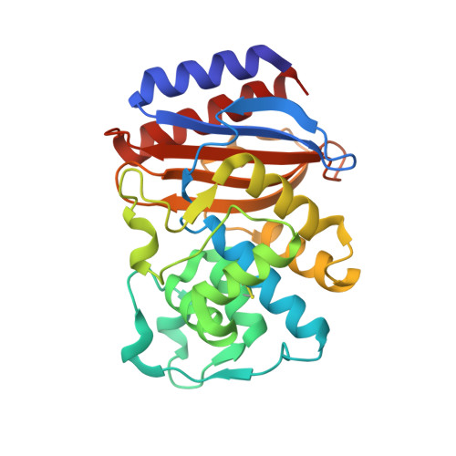
beta-Lactamases are bacterial enzymes which catalyse the hydrolysis of the beta-lactam ring of penicillins, cephalosporins and related compounds, thus inactivating these antibiotics. The crystal structure of the TEM1 beta-lactamase has been determined at 1.9 A resolution by the molecular-replacement method, using the atomic coordinates of two homologous beta-lactamase refined structures which show about 36% strict identity in their amino-acid sequences and 1.96 A r.m.s. deviation between equivalent Calpha atoms. The TEM1 enzyme crystallizes in space group P2(1)2(1)2(1) and there is one molecule per asymmetric unit. The structure was refined by simulated annealing to an R-factor of 15.6% for 15 086 reflections with I >/= 2sigma(I) in the resolution range 5.0-1.9 A. The final crystallographic structure contains 263 amino-acid residues, one sulfate anion in the catalytic cleft and 135 water molecules per asymmetric unit. The folding is very similar to that of the other known class A beta-lactamases. It consists of two domains, the first is formed by a five-stranded beta-sheet covered by three alpha-helices on one face and one alpha-helix on the other, the second domain contains mainly alpha-helices. The catalytic cleft is located at the interface between the two domains. We also report the crystallographic study of the TEM S235A mutant. This mutation of an active-site residue specifically decreases the acylation rate of cephalosporins. This TEM S235A mutant crystallizes under the same conditions as the wild-type protein and its structure was refined at 2.0 A resolution with an R value of 17.6%. The major modification is the appearance of a water molecule near the mutated residue, which is incompatible with the OG 235 present in the wild-type enzyme, and causes very small perturbations in the interaction network in the active site.
- Centre d'Ingénierie des Protéines, Unité de Cristallographie, Université de Liège, Institut de Physique, Belgium.
Organizational Affiliation: