1EQZ

X-RAY STRUCTURE OF THE NUCLEOSOME CORE PARTICLE AT 2.5 A RESOLUTION


Experimental Data Snapshot

  • Method: X-RAY DIFFRACTION
  • Resolution: 2.50 Å
  • R-Value Free: 0.282 
  • R-Value Work: 0.219 
  • R-Value Observed: 0.219 

wwPDB Validation   3D Report Full Report


This is version 1.4 of the entry. See complete history


Literature

Asymmetries in the nucleosome core particle at 2.5 A resolution.

Harp, J.M.Hanson, B.L.Timm, D.E.Bunick, G.J.

(2000) Acta Crystallogr D Biol Crystallogr 56: 1513-1534

  • DOI: https://doi.org/10.1107/s0907444900011847
  • Primary Citation of Related Structures:  
    1EQZ

  • PubMed Abstract: 

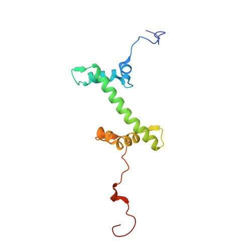
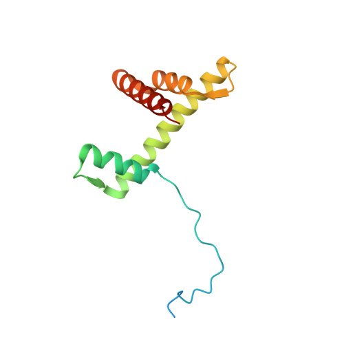
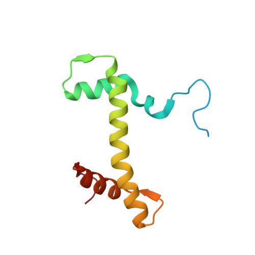
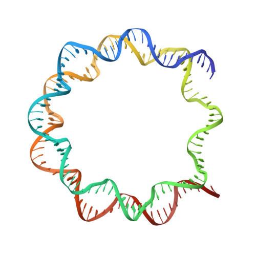
    The 2.5 A X-ray crystal structure of the nucleosome core particle presented here provides significant additions to the understanding of the nucleosome, the fundamental unit of chromatin structure. Extensions are made to the structure of the N-terminal histone tails and details are provided on hydration and ion binding. The structure is composed of twofold symmetric molecules, native chicken histone octamer cores and the DNA palindrome, which were expected to form a perfectly twofold symmetric nucleosome core particle. In fact, the result is asymmetric owing to the binding of the DNA to the protein surface and to the packing of the particles in the crystal lattice. An analysis is made of the asymmetries by comparisons both within the nucleosome core particle and to the structure of the histone octamer core of the nucleosome.


  • Organizational Affiliation

    University of Tennessee/Oak Ridge Graduate Program for Genome Sciences and Technology, Oak Ridge National Laboratory, Oak Ridge, TN 37831-8080, USA.


Macromolecules

Find similar proteins by:  (by identity cutoff)  |  3D Structure
Entity ID: 2
MoleculeChains Sequence LengthOrganismDetailsImage
PROTEIN (HISTONE H2A)C [auth A],
G [auth E]
129Gallus gallusMutation(s): 0 
UniProt
Find proteins for P02263 (Gallus gallus)
Explore P02263 
Go to UniProtKB:  P02263
Entity Groups  
Sequence Clusters30% Identity50% Identity70% Identity90% Identity95% Identity100% Identity
UniProt GroupP02263
Sequence Annotations
Expand
  • Reference Sequence
Find similar proteins by:  (by identity cutoff)  |  3D Structure
Entity ID: 3
MoleculeChains Sequence LengthOrganismDetailsImage
PROTEIN (HISTONE H2B)D [auth B],
H [auth F]
126Gallus gallusMutation(s): 0 
UniProt
Find proteins for P0C1H5 (Gallus gallus)
Explore P0C1H5 
Go to UniProtKB:  P0C1H5
Entity Groups  
Sequence Clusters30% Identity50% Identity70% Identity90% Identity95% Identity100% Identity
UniProt GroupP0C1H5
Sequence Annotations
Expand
  • Reference Sequence
Find similar proteins by:  (by identity cutoff)  |  3D Structure
Entity ID: 4
MoleculeChains Sequence LengthOrganismDetailsImage
PROTEIN (HISTONE H3)E [auth C],
I [auth G]
136Gallus gallusMutation(s): 0 
UniProt
Find proteins for P84229 (Gallus gallus)
Explore P84229 
Go to UniProtKB:  P84229
Entity Groups  
Sequence Clusters30% Identity50% Identity70% Identity90% Identity95% Identity100% Identity
UniProt GroupP84229
Sequence Annotations
Expand
  • Reference Sequence
Find similar proteins by:  (by identity cutoff)  |  3D Structure
Entity ID: 5
MoleculeChains Sequence LengthOrganismDetailsImage
PROTEIN (HISTONE H4)F [auth D],
J [auth H]
103Gallus gallusMutation(s): 0 
UniProt
Find proteins for P62801 (Gallus gallus)
Explore P62801 
Go to UniProtKB:  P62801
Entity Groups  
Sequence Clusters30% Identity50% Identity70% Identity90% Identity95% Identity100% Identity
UniProt GroupP62801
Sequence Annotations
Expand
  • Reference Sequence
Find similar nucleic acids by:  (by identity cutoff)  |  3D Structure
Entity ID: 1
MoleculeChains LengthOrganismImage
146 NUCLEOTIDES LONG DNAA [auth I],
B [auth J]
146N/A
Sequence Annotations
Expand
  • Reference Sequence
Small Molecules
Ligands 4 Unique
IDChains Name / Formula / InChI Key2D Diagram3D Interactions
CAC
Query on CAC

Download Ideal Coordinates CCD File 
IA [auth G]CACODYLATE ION
C2 H6 As O2
OGGXGZAMXPVRFZ-UHFFFAOYSA-M
MN
Query on MN

Download Ideal Coordinates CCD File 
AA [auth J]
BA [auth J]
CA [auth J]
DA [auth J]
EA [auth J]
AA [auth J],
BA [auth J],
CA [auth J],
DA [auth J],
EA [auth J],
FA [auth J],
L [auth I],
M [auth I],
N [auth I],
O [auth I],
P [auth I],
Q [auth I],
R [auth I],
S [auth I],
T [auth I]
MANGANESE (II) ION
Mn
WAEMQWOKJMHJLA-UHFFFAOYSA-N
K
Query on K

Download Ideal Coordinates CCD File 
K [auth I]
U [auth J]
V [auth J]
W [auth J]
X [auth J]
K [auth I],
U [auth J],
V [auth J],
W [auth J],
X [auth J],
Y [auth J],
Z [auth J]
POTASSIUM ION
K
NPYPAHLBTDXSSS-UHFFFAOYSA-N
CL
Query on CL

Download Ideal Coordinates CCD File 
GA [auth A],
HA [auth E]
CHLORIDE ION
Cl
VEXZGXHMUGYJMC-UHFFFAOYSA-M
Experimental Data & Validation

Experimental Data

  • Method: X-RAY DIFFRACTION
  • Resolution: 2.50 Å
  • R-Value Free: 0.282 
  • R-Value Work: 0.219 
  • R-Value Observed: 0.219 
  • Space Group: P 21 21 21
Unit Cell:
Length ( Å )Angle ( ˚ )
a = 105.28α = 90
b = 109.71β = 90
c = 181.12γ = 90
Software Package:
Software NamePurpose
SCALEPACKdata scaling
CCP4model building
CNSrefinement
CCP4phasing
CNSphasing

Structure Validation

View Full Validation Report


Entry History 

Deposition Data

Revision History  (Full details and data files)

  • Version 1.0: 2000-04-17
    Type: Initial release
  • Version 1.1: 2008-04-27
    Changes: Version format compliance
  • Version 1.2: 2011-07-13
    Changes: Non-polymer description, Version format compliance
  • Version 1.3: 2017-10-04
    Changes: Refinement description
  • Version 1.4: 2023-08-09
    Changes: Data collection, Database references, Derived calculations, Refinement description