1EHC | pdb_00001ehc

STRUCTURE OF SIGNAL TRANSDUCTION PROTEIN CHEY


Experimental Data Snapshot

  • Method: X-RAY DIFFRACTION
  • Resolution: 2.26 Å
  • R-Value Work: 
    0.143 (Depositor), 0.153 (DCC) 

Starting Model: experimental
View more details

wwPDB Validation 3D Report Full Report

Validation slider image for 1EHC

This is version 1.5 of the entry. See complete history

Literature

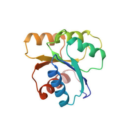
Uncoupled phosphorylation and activation in bacterial chemotaxis. The 2.3 A structure of an aspartate to lysine mutant at position 13 of CheY.

Jiang, M.Bourret, R.B.Simon, M.I.Volz, K.

(1997) J Biological Chem 272: 11850-11855

  • DOI: https://doi.org/10.1074/jbc.272.18.11850
  • Primary Citation Related Structures: 
    1EHC

  • PubMed Abstract: 

    An aspartate to lysine mutation at position 13 of the chemotaxis regulatory protein CheY causes a constitutive tumbly phenotype when expressed at high copy number in vivo even though the mutant protein is not phosphorylatable. These properties suggest that the D13K mutant adopts the active, signaling conformation of CheY independent of phosphorylation, so knowledge of its structure could explain the activation mechanism of CheY. The x-ray crystallographic structure of the CheY D13K mutant has been solved and refined at 2.3 A resolution to an R-factor of 14.3%. The mutant molecule shows no significant differences in backbone conformation when compared with the wild-type, Mg2+-free structure, but there are localized changes within the active site. The side chain of lysine 13 blocks access to the active site, whereas its epsilon-amino group has no bonding interactions with other groups in the region. Also in the active site, the bond between lysine 109 and aspartate 57 is weakened, and the solvent structure is perturbed. Although the D13K mutant has the inactive conformation in the crystalline form, rearrangements in the active site appear to weaken the overall structure of that region, potentially creating a metastable state of the molecule. If a conformational change is required for signaling by CheY D13K, then it most likely proceeds dynamically, in solution.


  • Organizational Affiliation
    • Department of Microbiology and Immunology, University of Illinois, Chicago, Illinois 60612, USA.

Macromolecule Content 

  • Total Structure Weight: 14.09 kDa 
  • Atom Count: 1,108 
  • Modeled Residue Count: 128 
  • Deposited Residue Count: 128 
  • Unique protein chains: 1

Macromolecules

Find similar proteins by:|  3D Structure
Entity ID: 1
MoleculeChains  Sequence LengthOrganismDetailsImage
CHEY128Escherichia coli K-12Mutation(s): 1 
UniProt
Find proteins for P0AE67 (Escherichia coli (strain K12))
Explore P0AE67 
Go to UniProtKB:  P0AE67
Entity Groups
Sequence Clusters30% Identity50% Identity70% Identity90% Identity95% Identity100% Identity
UniProt GroupP0AE67
Sequence Annotations
Expand
Reference Sequence

Small Molecules

Ligands 1 Unique
IDChains Name / Formula / InChI Key2D Diagram3D Interactions
SO4

Query on SO4


Download:Ideal Coordinates CCD File
B [auth A]SULFATE ION
O4 S
QAOWNCQODCNURD-UHFFFAOYSA-L

Experimental Data & Validation

Experimental Data

  • Method: X-RAY DIFFRACTION
  • Resolution: 2.26 Å
  • R-Value Work:  0.143 (Depositor), 0.153 (DCC) 
Space Group: P 21 21 21
Unit Cell:
Length ( Å )Angle ( ˚ )
a = 45.85α = 90
b = 47.03β = 90
c = 54.04γ = 90
Software Package:
Software NamePurpose
PROFFTrefinement
XENGENdata reduction
XENGENdata scaling

Structure Validation

View Full Validation Report


Entry History 

Deposition Data

Revision History  (Full details and data files)

  • Version 1.0: 1997-05-15
    Type: Initial release
  • Version 1.1: 2008-03-24
    Changes: Version format compliance
  • Version 1.2: 2011-07-13
    Changes: Source and taxonomy, Version format compliance
  • Version 1.3: 2021-11-03
    Changes: Database references, Derived calculations
  • Version 1.4: 2024-02-07
    Changes: Data collection
  • Version 1.5: 2026-03-04
    Changes: Refinement description, Structure summary