1EGJ | pdb_00001egj

DOMAIN 4 OF THE BETA COMMON CHAIN IN COMPLEX WITH AN ANTIBODY


Experimental Data Snapshot

  • Method: X-RAY DIFFRACTION
  • Resolution: 2.80 Å
  • R-Value Free: 
    0.288 (Depositor) 
  • R-Value Work: 
    0.228 (Depositor) 
  • R-Value Observed: 
    0.220 (Depositor) 

wwPDB Validation 3D Report Full Report

Validation slider image for 1EGJ

This is version 1.5 of the entry. See complete history

Literature

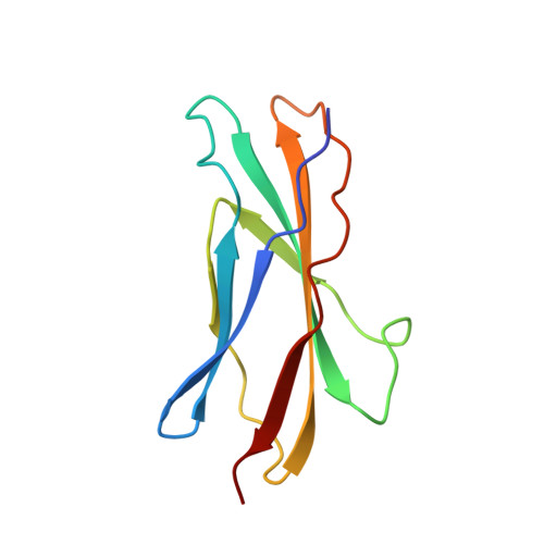
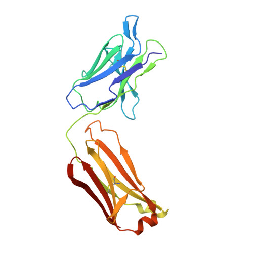
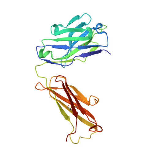
Structure of the activation domain of the GM-CSF/IL-3/IL-5 receptor common beta-chain bound to an antagonist.

Rossjohn, J.McKinstry, W.J.Woodcock, J.M.McClure, B.J.Hercus, T.R.Parker, M.W.Lopez, A.F.Bagley, C.J.

(2000) Blood 95: 2491-2498

  • Primary Citation Related Structures: 
    1EGJ

  • PubMed Abstract: 

    Heterodimeric cytokine receptors generally consist of a major cytokine-binding subunit and a signaling subunit. The latter can transduce signals by more than 1 cytokine, as exemplified by the granulocyte-macrophage colony-stimulating factor (GM-CSF), interleukin-2 (IL-2), and IL-6 receptor systems. However, often the signaling subunits in isolation are unable to bind cytokines, a fact that has made it more difficult to obtain structural definition of their ligand-binding sites. This report details the crystal structure of the ligand-binding domain of the GM-CSF/IL-3/IL-5 receptor beta-chain (beta(c)) signaling subunit in complex with the Fab fragment of the antagonistic monoclonal antibody, BION-1. This is the first single antagonist of all 3 known eosinophil-producing cytokines, and it is therefore capable of regulating eosinophil-related diseases such as asthma. The structure reveals a fibronectin type III domain, and the antagonist-binding site involves major contributions from the loop between the B and C strands and overlaps the cytokine-binding site. Furthermore, tyrosine(421) (Tyr(421)), a key residue involved in receptor activation, lies in the neighboring loop between the F and G strands, although it is not immediately adjacent to the cytokine-binding residues in the B-C loop. Interestingly, functional experiments using receptors mutated across these loops demonstrate that they are cooperatively involved in full receptor activation. The experiments, however, reveal subtle differences between the B-C loop and Tyr(421), which is suggestive of distinct functional roles. The elucidation of the structure of the ligand-binding domain of beta(c) also suggests how different cytokines recognize a single receptor subunit, which may have implications for homologous receptor systems. (Blood. 2000;95:2491-2498)


  • Organizational Affiliation
    • Ian Potter Foundation Protein Crystallography Laboratory, St. Vincent's Institute of Medical Research, Fitzroy, Victoria, Australia.

Macromolecule Content 

  • Total Structure Weight: 59.18 kDa 
  • Atom Count: 4,304 
  • Modeled Residue Count: 536 
  • Deposited Residue Count: 536 
  • Unique protein chains: 3

Macromolecules

Find similar proteins by:|  3D Structure
Entity ID: 1
MoleculeChains  Sequence LengthOrganismDetailsImage
CYTOKINE RECEPTOR COMMON BETA CHAIN PRECURSOR101Homo sapiensMutation(s): 0 
UniProt & NIH Common Fund Data Resources
Find proteins for P32927 (Homo sapiens)
Explore P32927 
Go to UniProtKB:  P32927
PHAROS:  P32927
GTEx:  ENSG00000100368 
Entity Groups
Sequence Clusters30% Identity50% Identity70% Identity90% Identity95% Identity100% Identity
UniProt GroupP32927
Sequence Annotations
Expand
Reference Sequence
Find similar proteins by:|  3D Structure
Entity ID: 2
MoleculeChains  Sequence LengthOrganismDetailsImage
ANTIBODY (LIGHT CHAIN)B [auth L]215Mus musculusMutation(s): 0 
UniProt
Find proteins for P01661 (Mus musculus)
Explore P01661 
Go to UniProtKB:  P01661
Entity Groups
Sequence Clusters30% Identity50% Identity70% Identity90% Identity95% Identity100% Identity
UniProt GroupP01661
Glycosylation
Glycosylation Sites: 1Go to GlyGen: P01661-1
Sequence Annotations
Expand
Reference Sequence
Find similar proteins by:|  3D Structure
Entity ID: 3
MoleculeChains  Sequence LengthOrganismDetailsImage
ANTIBODY (HEAVY CHAIN)C [auth H]220Mus musculusMutation(s): 0 
UniProt
Find proteins for P01865 (Mus musculus)
Explore P01865 
Go to UniProtKB:  P01865
Entity Groups
Sequence Clusters30% Identity50% Identity70% Identity90% Identity95% Identity100% Identity
UniProt GroupP01865
Sequence Annotations
Expand
Reference Sequence

Small Molecules

Ligands 1 Unique
IDChains Name / Formula / InChI Key2D Diagram3D Interactions
NAG

Query on NAG


Download:Ideal Coordinates CCD File
D [auth L]2-acetamido-2-deoxy-beta-D-glucopyranose
C8 H15 N O6
OVRNDRQMDRJTHS-FMDGEEDCSA-N

Experimental Data & Validation

Experimental Data

  • Method: X-RAY DIFFRACTION
  • Resolution: 2.80 Å
  • R-Value Free:  0.288 (Depositor) 
  • R-Value Work:  0.228 (Depositor) 
  • R-Value Observed: 0.220 (Depositor) 
Space Group: P 41 21 2
Unit Cell:
Length ( Å )Angle ( ˚ )
a = 77.61α = 90
b = 77.61β = 90
c = 296.25γ = 90
Software Package:
Software NamePurpose
DENZOdata reduction
SCALEPACKdata scaling
AMoREphasing
CNSrefinement

Structure Validation

View Full Validation Report


Entry History 

Deposition Data

Revision History  (Full details and data files)

  • Version 1.0: 2001-02-15
    Type: Initial release
  • Version 1.1: 2008-04-27
    Changes: Version format compliance
  • Version 1.2: 2011-07-13
    Changes: Non-polymer description, Version format compliance
  • Version 1.3: 2018-01-31
    Changes: Experimental preparation
  • Version 1.4: 2020-07-29
    Type: Remediation
    Reason: Carbohydrate remediation
    Changes: Data collection, Database references, Derived calculations, Structure summary
  • Version 1.5: 2024-10-09
    Changes: Data collection, Database references, Structure summary