1EFX | pdb_00001efx

STRUCTURE OF A COMPLEX BETWEEN THE HUMAN NATURAL KILLER CELL RECEPTOR KIR2DL2 AND A CLASS I MHC LIGAND HLA-CW3


Experimental Data Snapshot

  • Method: X-RAY DIFFRACTION
  • Resolution: 3.00 Å
  • R-Value Free: 
    0.294 (Depositor) 
  • R-Value Work: 
    0.231 (Depositor) 
  • R-Value Observed: 
    0.209 (Depositor) 

Starting Models: experimental
View more details

wwPDB Validation 3D Report Full Report

Validation slider image for 1EFX

This is version 1.4 of the entry. See complete history

Literature

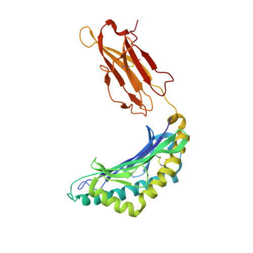
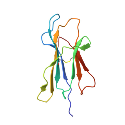
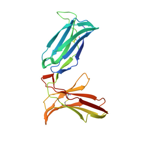
Crystal structure of an NK cell immunoglobulin-like receptor in complex with its class I MHC ligand.

Boyington, J.C.Motyka, S.A.Schuck, P.Brooks, A.G.Sun, P.D.

(2000) Nature 405: 537-543

  • DOI: https://doi.org/10.1038/35014520
  • Primary Citation Related Structures: 
    1EFX

  • PubMed Abstract: 

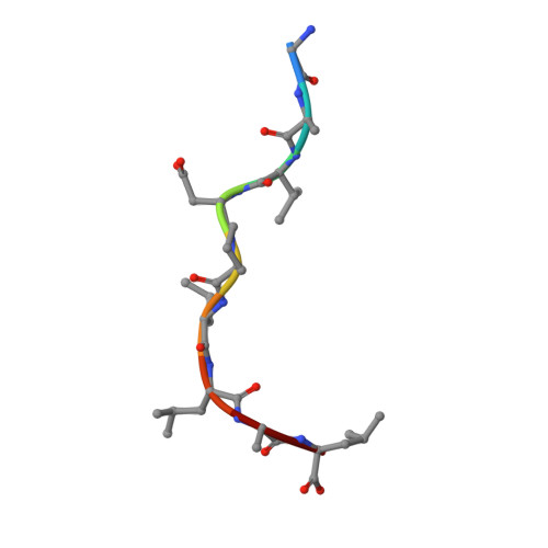
    Target cell lysis is regulated by natural killer (NK) cell receptors that recognize class I MHC molecules. Here we report the crystal structure of the human immunoglobulin-like NK cell receptor KIR2DL2 in complex with its class I ligand HLA-Cw3 and peptide. KIR binds in a nearly orthogonal orientation across the alpha1 and alpha2 helices of Cw3 and directly contacts positions 7 and 8 of the peptide. No significant conformational changes in KIR occur on complex formation. The receptor footprint on HLA overlaps with but is distinct from that of the T-cell receptor. Charge complementarity dominates the KIR/HLA interface and mutations that disrupt interface salt bridges substantially diminish binding. Most contacts in the complex are between KIR and conserved HLA-C residues, but a hydrogen bond between Lys 44 of KIR2DL2 and Asn 80 of Cw3 confers the allotype specificity. KIR contact requires position 8 of the peptide to be a residue smaller than valine. A second KIR/HLA interface produced an ordered receptor-ligand aggregation in the crystal which may resemble receptor clustering during immune synapse formation.


  • Organizational Affiliation
    • Structural Biology Section, Laboratory of Immunogenetics, National Institute of Allergy and Infectious Diseases, National Institutes of Health, Rockville, Maryland 20852, USA.

Macromolecule Content 

  • Total Structure Weight: 89.03 kDa 
  • Atom Count: 6,411 
  • Modeled Residue Count: 781 
  • Deposited Residue Count: 787 
  • Unique protein chains: 4

Macromolecules

Find similar proteins by:|  3D Structure
Entity ID: 1
MoleculeChains  Sequence LengthOrganismDetailsImage
HLA-CW3 (HEAVY CHAIN)278Homo sapiensMutation(s): 0 
UniProt & NIH Common Fund Data Resources
Find proteins for P10321 (Homo sapiens)
Explore P10321 
Go to UniProtKB:  P10321
PHAROS:  P10321
GTEx:  ENSG00000204525 
Entity Groups
Sequence Clusters30% Identity50% Identity70% Identity90% Identity95% Identity100% Identity
UniProt GroupP10321
Sequence Annotations
Expand
Reference Sequence
Find similar proteins by:|  3D Structure
Entity ID: 2
MoleculeChains  Sequence LengthOrganismDetailsImage
BETA-2-MICROGLOBULIN100Homo sapiensMutation(s): 0 
UniProt & NIH Common Fund Data Resources
Find proteins for P61769 (Homo sapiens)
Explore P61769 
Go to UniProtKB:  P61769
PHAROS:  P61769
GTEx:  ENSG00000166710 
Entity Groups
Sequence Clusters30% Identity50% Identity70% Identity90% Identity95% Identity100% Identity
UniProt GroupP61769
Sequence Annotations
Expand
Reference Sequence
Find similar proteins by:  Sequence   |   3D Structure  
Entity ID: 3
MoleculeChains  Sequence LengthOrganismDetailsImage
PEPTIDE FROM IMPORTIN ALPHA-29N/AMutation(s): 0 
UniProt & NIH Common Fund Data Resources
Find proteins for P52292 (Homo sapiens)
Explore P52292 
Go to UniProtKB:  P52292
PHAROS:  P52292
GTEx:  ENSG00000182481 
Entity Groups
UniProt GroupP52292
Sequence Annotations
Expand
Reference Sequence
Find similar proteins by:|  3D Structure
Entity ID: 4
MoleculeChains  Sequence LengthOrganismDetailsImage
NATURAL KILLER CELL RECEPTOR KIR2DL2
D, E
200Homo sapiensMutation(s): 0 
UniProt
Find proteins for P43627 (Homo sapiens)
Explore P43627 
Go to UniProtKB:  P43627
Entity Groups
Sequence Clusters30% Identity50% Identity70% Identity90% Identity95% Identity100% Identity
UniProt GroupP43627
Sequence Annotations
Expand
Reference Sequence

Experimental Data & Validation

Experimental Data

  • Method: X-RAY DIFFRACTION
  • Resolution: 3.00 Å
  • R-Value Free:  0.294 (Depositor) 
  • R-Value Work:  0.231 (Depositor) 
  • R-Value Observed: 0.209 (Depositor) 
Space Group: P 21 21 21
Unit Cell:
Length ( Å )Angle ( ˚ )
a = 68.54α = 90
b = 90.33β = 90
c = 207.27γ = 90
Software Package:
Software NamePurpose
DENZOdata reduction
SCALEPACKdata scaling
AMoREphasing
CNSrefinement

Structure Validation

View Full Validation Report


Entry History 

Deposition Data

Revision History  (Full details and data files)

  • Version 1.0: 2000-06-14
    Type: Initial release
  • Version 1.1: 2008-04-27
    Changes: Version format compliance
  • Version 1.2: 2011-07-13
    Changes: Version format compliance
  • Version 1.3: 2023-08-09
    Changes: Data collection, Database references, Refinement description
  • Version 1.4: 2024-11-13
    Changes: Structure summary