1E91 | pdb_00001e91

Structure of the complex of the Mad1-Sin3B interaction domains


Experimental Data Snapshot

  • Method: SOLUTION NMR
  • Conformers Calculated: 100 
  • Conformers Submitted: 20 
  • Selection Criteria: 30 STRUCTURES WITH NO RESTRAINT VIOLATIONS, 20 LOWEST ENERGY STRUCTURES FINALLY SELECTED 

wwPDB Validation 3D Report Full Report

Validation slider image for 1E91

This is version 1.4 of the entry. See complete history

Literature

The MAD1-Sin3B Interaction Involves a Novel Helical Fold

Spronk, C.A.E.M.Tessari, M.Kaan, A.M.Jansen, J.F.A.Vermeulen, M.Stunnenberg, H.G.Vuister, G.W.

(2000) Nat Struct Biol 7: 1100-1104

  • DOI: https://doi.org/10.1038/81944
  • Primary Citation Related Structures: 
    1E91

  • PubMed Abstract: 

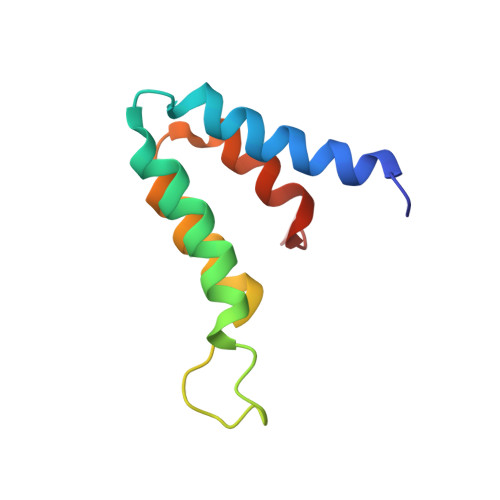
    Sin3A or Sin3B are components of a corepressor complex that mediates repression by transcription factors such as the helix-loop-helix proteins Mad and Mxi. Members of the Mad/Mxi family of repressors play important roles in the transition between proliferation and differentiation by down-regulating the expression of genes that are activated by the proto-oncogene product Myc. Here, we report the solution structure of the second paired amphipathic helix (PAH) domain (PAH2) of Sin3B in complex with a peptide comprising the N-terminal region of Mad1. This complex exhibits a novel interaction fold for which we propose the name 'wedged helical bundle'. Four alpha-helices of PAH2 form a hydrophobic cleft that accommodates an amphipathic Mad1 alpha-helix. Our data further show that, upon binding Mad1, secondary structure elements of PAH2 are stabilized. The PAH2-Mad1 structure provides the basis for determining the principles of protein interaction and selectivity involving PAH domains.


  • Organizational Affiliation
    • Department of Biophysical Chemistry, NSR Center, University of Nijmegen, The Netherlands.

Macromolecule Content 

  • Total Structure Weight: 11.62 kDa 
  • Atom Count: 820 
  • Modeled Residue Count: 98 
  • Deposited Residue Count: 98 
  • Unique protein chains: 2

Macromolecules

Find similar proteins by:|  3D Structure
Entity ID: 1
MoleculeChains  Sequence LengthOrganismDetailsImage
PAIRED AMPHIPATHIC HELIX PROTEIN SIN3B85Mus musculusMutation(s): 0 
Gene Names: SIN3B
UniProt
Find proteins for Q62141 (Mus musculus)
Explore Q62141 
Go to UniProtKB:  Q62141
Entity Groups
Sequence Clusters30% Identity50% Identity70% Identity90% Identity95% Identity100% Identity
UniProt GroupQ62141
Sequence Annotations
Expand
Reference Sequence
Find similar proteins by:  Sequence   |   3D Structure  
Entity ID: 2
MoleculeChains  Sequence LengthOrganismDetailsImage
MAD PROTEIN (MAX DIMERIZER)13Homo sapiensMutation(s): 0 
UniProt & NIH Common Fund Data Resources
Find proteins for Q05195 (Homo sapiens)
Explore Q05195 
Go to UniProtKB:  Q05195
PHAROS:  Q05195
GTEx:  ENSG00000059728 
Entity Groups
Sequence Clusters30% Identity50% Identity70% Identity90% Identity95% Identity100% Identity
UniProt GroupQ05195
Sequence Annotations
Expand
Reference Sequence

Experimental Data & Validation

Experimental Data

  • Method: SOLUTION NMR
  • Conformers Calculated: 100 
  • Conformers Submitted: 20 
  • Selection Criteria: 30 STRUCTURES WITH NO RESTRAINT VIOLATIONS, 20 LOWEST ENERGY STRUCTURES FINALLY SELECTED 

Structure Validation

View Full Validation Report


Entry History 

Deposition Data

Revision History  (Full details and data files)

  • Version 1.0: 2000-11-20
    Type: Initial release
  • Version 1.1: 2011-05-08
    Changes: Version format compliance
  • Version 1.2: 2011-07-13
    Changes: Version format compliance
  • Version 1.3: 2018-01-17
    Changes: Database references, Source and taxonomy
  • Version 1.4: 2024-05-15
    Changes: Data collection, Database references, Other