1E5Q

Ternary complex of saccharopine reductase from Magnaporthe grisea, NADPH and saccharopine


Experimental Data Snapshot

  • Method: X-RAY DIFFRACTION
  • Resolution: 2.10 Å
  • R-Value Free: 0.228 
  • R-Value Work: 0.207 
  • R-Value Observed: 0.207 

wwPDB Validation   3D Report Full Report


Ligand Structure Quality Assessment 


This is version 1.6 of the entry. See complete history


Literature

Crystal Structure of Saccharopine Reductase from Magnaporthe Grisea, an Enzyme of the Alpha-Aminoadipate Pathway of Lysine Biosynthesis

Johansson, E.Steffens, J.J.Lindqvist, Y.Schneider, G.

(2000) Structure 8: 1037

  • DOI: https://doi.org/10.1016/s0969-2126(00)00512-8
  • Primary Citation of Related Structures:  
    1E5L, 1E5Q, 1FF9

  • PubMed Abstract: 

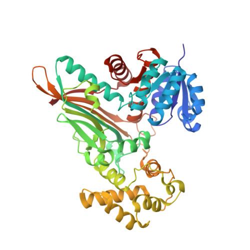
    The biosynthesis of the essential amino acid lysine in higher fungi and cyanobacteria occurs via the alpha-aminoadipate pathway, which is completely different from the lysine biosynthetic pathway found in plants and bacteria. The penultimate reaction in the alpha-aminoadipate pathway is catalysed by NADPH-dependent saccharopine reductase. We set out to determine the structure of this enzyme as a first step in exploring the structural biology of fungal lysine biosynthesis. We have determined the three-dimensional structure of saccharopine reductase from the plant pathogen Magnaporthe grisea in its apo form to 2.0 A resolution and as a ternary complex with NADPH and saccharopine to 2.1 A resolution. Saccharopine reductase is a homodimer, and each subunit consists of three domains, which are not consecutive in amino acid sequence. Domain I contains a variant of the Rossmann fold that binds NADPH. Domain II folds into a mixed seven-stranded beta sheet flanked by alpha helices and is involved in substrate binding and dimer formation. Domain III is all-helical. The structure analysis of the ternary complex reveals a large movement of domain III upon ligand binding. The active site is positioned in a cleft between the NADPH-binding domain and the second alpha/beta domain. Saccharopine is tightly bound to the enzyme via a number of hydrogen bonds to invariant amino acid residues. On the basis of the structure of the ternary complex of saccharopine reductase, an enzymatic mechanism is proposed that includes the formation of a Schiff base as a key intermediate. Despite the lack of overall sequence homology, the fold of saccharopine reductase is similar to that observed in some enzymes of the diaminopimelate pathway of lysine biosynthesis in bacteria. These structural similarities suggest an evolutionary relationship between two different major families of amino acid biosynthetic pathway, the glutamate and aspartate families.


  • Organizational Affiliation

    Department of Medical Biochemistry and Biophysics Karolinska Institutet S-171 77, Stockholm, Sweden.


Macromolecules
Find similar proteins by:  (by identity cutoff)  |  3D Structure
Entity ID: 1
MoleculeChains Sequence LengthOrganismDetailsImage
Saccharopine dehydrogenase [NADP(+), L-glutamate-forming]
A, B, C, D, E
A, B, C, D, E, F, G, H
450Pyricularia griseaMutation(s): 0 
Gene Names: LYS3MGG_08564
EC: 1.5.1.10
UniProt
Find proteins for Q9P4R4 (Pyricularia oryzae (strain 70-15 / ATCC MYA-4617 / FGSC 8958))
Explore Q9P4R4 
Go to UniProtKB:  Q9P4R4
Entity Groups  
Sequence Clusters30% Identity50% Identity70% Identity90% Identity95% Identity100% Identity
UniProt GroupQ9P4R4
Sequence Annotations
Expand
  • Reference Sequence
Small Molecules
Ligands 2 Unique
IDChains Name / Formula / InChI Key2D Diagram3D Interactions
NDP
Query on NDP

Download Ideal Coordinates CCD File 
I [auth A]
K [auth B]
M [auth C]
O [auth D]
Q [auth E]
I [auth A],
K [auth B],
M [auth C],
O [auth D],
Q [auth E],
S [auth F],
U [auth G],
W [auth H]
NADPH DIHYDRO-NICOTINAMIDE-ADENINE-DINUCLEOTIDE PHOSPHATE
C21 H30 N7 O17 P3
ACFIXJIJDZMPPO-NNYOXOHSSA-N
SHR
Query on SHR

Download Ideal Coordinates CCD File 
J [auth A]
L [auth B]
N [auth C]
P [auth D]
R [auth E]
J [auth A],
L [auth B],
N [auth C],
P [auth D],
R [auth E],
T [auth F],
V [auth G],
X [auth H]
N-(5-AMINO-5-CARBOXYPENTYL)GLUTAMIC ACID
C11 H20 N2 O6
ZDGJAHTZVHVLOT-YUMQZZPRSA-N
Experimental Data & Validation

Experimental Data

  • Method: X-RAY DIFFRACTION
  • Resolution: 2.10 Å
  • R-Value Free: 0.228 
  • R-Value Work: 0.207 
  • R-Value Observed: 0.207 
  • Space Group: P 1 21 1
Unit Cell:
Length ( Å )Angle ( ˚ )
a = 86.643α = 90
b = 205.809β = 100.05
c = 125.599γ = 90
Software Package:
Software NamePurpose
CNSrefinement
DENZOdata reduction
SCALEPACKdata scaling
EPMRphasing

Structure Validation

View Full Validation Report


Ligand Structure Quality Assessment 


Entry History 

Deposition Data

Revision History  (Full details and data files)

  • Version 1.0: 2000-12-03
    Type: Initial release
  • Version 1.1: 2011-05-08
    Changes: Version format compliance
  • Version 1.2: 2011-07-13
    Changes: Version format compliance
  • Version 1.3: 2018-06-13
    Changes: Data collection, Database references, Source and taxonomy, Structure summary
  • Version 1.4: 2019-07-10
    Changes: Data collection
  • Version 1.5: 2019-07-24
    Changes: Data collection
  • Version 1.6: 2023-12-13
    Changes: Data collection, Database references, Derived calculations, Other, Refinement description