1E31 | pdb_00001e31

SURVIVIN DIMER H. SAPIENS


Experimental Data Snapshot

  • Method: X-RAY DIFFRACTION
  • Resolution: 2.71 Å
  • R-Value Free: 
    0.270 (Depositor) 
  • R-Value Work: 
    0.235 (Depositor) 
  • R-Value Observed: 
    0.235 (Depositor) 

wwPDB Validation 3D Report Full Report

Validation slider image for 1E31

This is version 1.3 of the entry. See complete history

Literature

Crystal Structure of Human Survivin Reveals a Bow Tie-Shaped Dimer with Two Unusual Alpha-Helical Extensions

Chantalat, L.Skoufias, D.A.Kleman, J.P.Jung, B.Dideberg, O.Margolis, R.L.

(2000) Mol Cell 6: 183

  • Primary Citation Related Structures: 
    1E31

  • PubMed Abstract: 

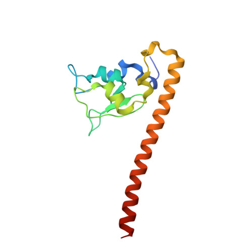
    Survivin is a mitotic spindle-associated protein involved in linking mitotic spindle function to activation of apoptosis in mammalian cells. The structure of the full-length human survivin has been determined by X-ray crystallography to 2.7 A. Strikingly, the structure forms a very unusual bow tie-shaped dimer. It does not dimerize through a C-terminal coiled-coil, contrary to sequence analysis prediction. The C-terminal helices contain hydrophobic clusters with the potential for protein-protein interactions. The unusual shape and dimensions of survivin suggest it serves an adaptor function through its alpha-helical extensions.


  • Organizational Affiliation
    • Laboratoire de Cristallographie Macromoléculaire, Institut de Biologie Structurale Jean-Pierre Ebel, CEA-CNRS, Grenoble, France.

Macromolecule Content 

  • Total Structure Weight: 33.02 kDa 
  • Atom Count: 2,243 
  • Modeled Residue Count: 274 
  • Deposited Residue Count: 284 
  • Unique protein chains: 1

Macromolecules

Find similar proteins by:|  3D Structure
Entity ID: 1
MoleculeChains  Sequence LengthOrganismDetailsImage
APOPTOSIS INHIBITOR SURVIVIN
A, B
142Homo sapiensMutation(s): 0 
UniProt & NIH Common Fund Data Resources
Find proteins for O15392 (Homo sapiens)
Explore O15392 
Go to UniProtKB:  O15392
PHAROS:  O15392
GTEx:  ENSG00000089685 
Entity Groups
Sequence Clusters30% Identity50% Identity70% Identity90% Identity95% Identity100% Identity
UniProt GroupO15392
Sequence Annotations
Expand
Reference Sequence

Experimental Data & Validation

Experimental Data

  • Method: X-RAY DIFFRACTION
  • Resolution: 2.71 Å
  • R-Value Free:  0.270 (Depositor) 
  • R-Value Work:  0.235 (Depositor) 
  • R-Value Observed: 0.235 (Depositor) 
Space Group: I 1 2 1
Unit Cell:
Length ( Å )Angle ( ˚ )
a = 82.22α = 90
b = 71.05β = 96.12
c = 89.45γ = 90
Software Package:
Software NamePurpose
CNSrefinement
DENZOdata reduction
SCALEPACKdata scaling
CNSphasing

Structure Validation

View Full Validation Report


Entry History 

Deposition Data

Revision History  (Full details and data files)

  • Version 1.0: 2001-01-03
    Type: Initial release
  • Version 1.1: 2011-05-08
    Changes: Version format compliance
  • Version 1.2: 2011-07-13
    Changes: Version format compliance
  • Version 1.3: 2024-05-08
    Changes: Data collection, Database references, Derived calculations