Isomorphous crystal structures of Escherichia coli dihydrofolate reductase complexed with folate, 5-deazafolate, and 5,10-dideazatetrahydrofolate: mechanistic implications.
Reyes, V.M., Sawaya, M.R., Brown, K.A., Kraut, J.(1995) Biochemistry 34: 2710-2723
- PubMed: 7873554 Search on PubMed
- DOI: https://doi.org/10.1021/bi00008a039
- Primary Citation Related Structures:
1DRH, 1DYH, 1DYI, 1DYJ - PubMed Abstract:
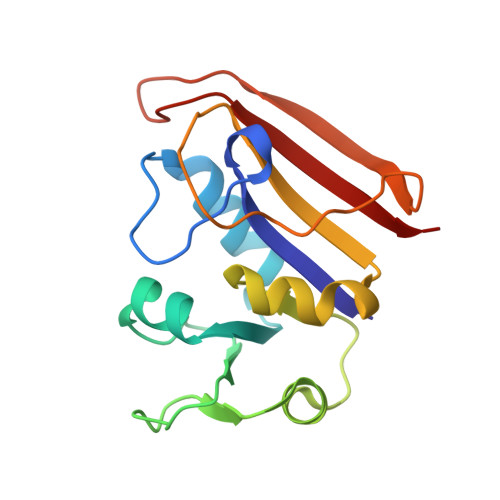
Crystal structures of Escherichia coli dihydrofolate reductase (ecDHFR, EC 1.5.1.3) in binary complexes with folate, 5-deazafolate (5dfol), and 5,10-dideazatetrahydrofolate (ddTHF) have been refined to R-factors of 13.7%, 14.9%, and 14.5%, respectively, all at 1.9 A. All three are isomorphous with a previously reported binary complex of ecDHFR with methotrexate (MTX), in space group P6(1), two molecules per asymmetric unit [Bolin, J. T., Filman, D. J., Matthews, D. A., Hamlin, R. C., & Kraut, J. (1982) J. Biol. Chem. 257, 13650-13662]. A hitherto unobserved water molecule is hydrogen bonded to the pteridine N5 and O4 in both molecules of the asymmetric unit of the folate complex (but not the 5dfol or ddTHF complexes), supporting the hypothesis that N5 protonation of bound substrate, an important step of the DHFR reaction, occurs by way of such a water molecule. There is no indication of a hydrogen bond between N8 of 5dfol and the backbone carbonyl of Ile-5, suggesting that the bacterial enzyme, unlike the human enzyme [Davies, J. F., II, Delcamp, T. J., Prendergast, N. J., Ashford, V. A., Freisheim, J. H., & Kraut, J. (1990) Biochemistry 29, 9467-9479], does not favor protonation at N8. Perhaps this explains why bacterial DHFR is much less effective than vertebrate DHFR in folate reduction. When the ecDHFR.NADPH complex (space group P3221; M. R. Sawaya, in preparation) is superimposed on the folate and 5dfol complexes, the distances from pteridine C6 to nicotinamide C4 were found to be 2.9 and 2.8 A, respectively, in close agreement with the theoretically calculated optimal distance in the transition state for hydride transfer [Wu, Y. D., & Houk, K. N. (1987) J. Am. Chem. Soc. 109, 906-908, 2226-2227]. In contrast to the planar ring system of folate or 5dfol, the reduced pteridine ring of ddTHF is severely puckered and bent toward the nicotinamide pocket, with the reduced pyridine ring assuming a half-chair type of conformation. This change in shape causes the pteridine ring to bind with O4 closer to Trp-22(N epsilon 1) by over 0.5 A, so that an invariant water molecule now bridges these two atoms with ideal hydrogen bonds. Furthermore, while the pABA rings of folate and 5dfol are nearly coincident and closer to the alpha C helix than to the alpha B helix, those of MTX and ddTHF are displaced along the binding crevice by approximately 1.1 and 0.6 A, respectively, and are equidistant from alpha B and alpha C.(ABSTRACT TRUNCATED AT 400 WORDS)
- Department of Chemistry and Biochemistry, University of California, San Diego, La Jolla 92093-0317.
Organizational Affiliation: