1DXJ

Structure of the chitinase from jack bean


Experimental Data Snapshot

  • Method: X-RAY DIFFRACTION
  • Resolution: 1.80 Å
  • R-Value Free: 0.222 
  • R-Value Work: 0.182 
  • R-Value Observed: 0.182 

wwPDB Validation   3D Report Full Report


This is version 1.3 of the entry. See complete history


Literature

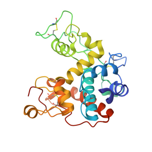
Structure of Jack Bean Chitinase

Hahn, M.Hennig, M.Schlesier, B.Hohne, W.

(2000) Acta Crystallogr D Biol Crystallogr 56: 1096

  • DOI: https://doi.org/10.1107/s090744490000857x
  • Primary Citation of Related Structures:  
    1DXJ

  • PubMed Abstract: 
  • The structure of jack bean chitinase was solved at 1.8 A resolution by molecular replacement. It is an alpha-helical protein with three disulfide bridges. The active site is related in structure to animal and viral lysozymes. However, unlike in lysozyme, the architecture of the active site suggests a single-step cleavage ...

    The structure of jack bean chitinase was solved at 1.8 A resolution by molecular replacement. It is an alpha-helical protein with three disulfide bridges. The active site is related in structure to animal and viral lysozymes. However, unlike in lysozyme, the architecture of the active site suggests a single-step cleavage. According to this mechanism, Glu68 is the proton donor and Glu90 assists in the reaction by moving towards the substrate and recruiting a water molecule that acts as the nucleophile. In this model, a water molecule was found in contact with Glu90 O(epsilon1) and Thr119 O(gamma) at a distance of 3.0 and 2.8 A, respectively. The model is in accordance with the observed inversion mechanism.


    Related Citations: 
    • Refined Structure of the Chitinase from Barley Seeds at 2.0 A Resolution
      Song, S.
      (1996) Acta Crystallogr D Biol Crystallogr 52: 289
    • Crystal Structure of an Endochitinase from Hordeum Vulgare L. Seeds
      Hartmonzingoready, E.J.
      (1993) J Mol Biol 229: 189

    Organizational Affiliation

    Institut für Biochemie, Charité, Humboldt-Universität, Monbijoustrasse 2, 10117 Berlin, Germany. michael.hahn@charite.de


Macromolecules
Find similar proteins by:  (by identity cutoff)  |  3D Structure
Entity ID: 1
MoleculeChainsSequence LengthOrganismDetailsImage
CLASS II CHITINASE242Canavalia ensiformisMutation(s): 0 
EC: 3.2.1.14
Entity Groups  
Sequence Clusters30% Identity50% Identity70% Identity90% Identity95% Identity100% Identity
Protein Feature View
Expand
  • Reference Sequence
Small Molecules
Ligands 1 Unique
IDChainsName / Formula / InChI Key2D Diagram3D Interactions
SO4
Query on SO4

Download Ideal Coordinates CCD File 
B [auth A]SULFATE ION
O4 S
QAOWNCQODCNURD-UHFFFAOYSA-L
 Ligand Interaction
Experimental Data & Validation

Experimental Data

  • Method: X-RAY DIFFRACTION
  • Resolution: 1.80 Å
  • R-Value Free: 0.222 
  • R-Value Work: 0.182 
  • R-Value Observed: 0.182 
  • Space Group: P 61
Unit Cell:
Length ( Å )Angle ( ˚ )
a = 67.69α = 90
b = 67.69β = 90
c = 110.21γ = 120
Software Package:
Software NamePurpose
X-PLORrefinement
XDSdata reduction
XDSdata scaling
X-PLORphasing

Structure Validation

View Full Validation Report


Entry History 

Deposition Data

Revision History  (Full details and data files)

  • Version 1.0: 2000-08-29
    Type: Initial release
  • Version 1.1: 2011-05-07
    Changes: Version format compliance
  • Version 1.2: 2011-07-13
    Changes: Version format compliance
  • Version 1.3: 2019-05-08
    Changes: Data collection, Experimental preparation, Other