1DVR

STRUCTURE OF A MUTANT ADENYLATE KINASE LIGATED WITH AN ATP-ANALOGUE SHOWING DOMAIN CLOSURE OVER ATP


Experimental Data Snapshot

  • Method: X-RAY DIFFRACTION
  • Resolution: 2.36 Å
  • R-Value Work: 0.191 
  • R-Value Observed: 0.191 

wwPDB Validation   3D Report Full Report


Ligand Structure Quality Assessment 


This is version 1.5 of the entry. See complete history


Literature

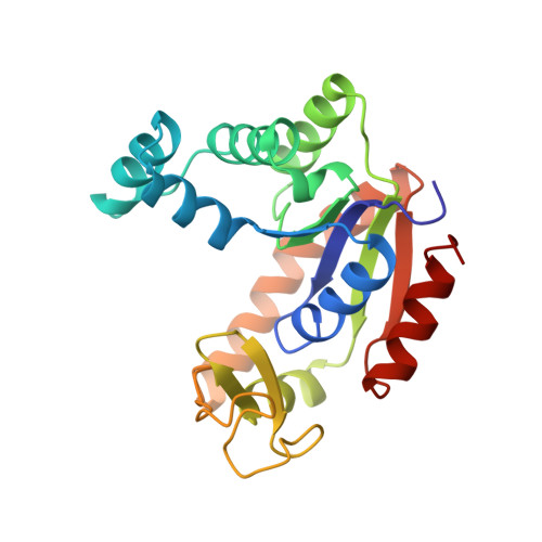
Structure of a mutant adenylate kinase ligated with an ATP-analogue showing domain closure over ATP.

Schlauderer, G.J.Proba, K.Schulz, G.E.

(1996) J Mol Biol 256: 223-227

  • DOI: https://doi.org/10.1006/jmbi.1996.0080
  • Primary Citation of Related Structures:  
    1DVR

  • PubMed Abstract: 

    Structural studies on unligated and ligated adenylate kinases have shown that two domains, LID and NMPbind, close over the bound substrates, ATP and AMP, respectively. These motions can be, but need not be independent from each other. Up to now, the known structures display only the states "both domains open", "both closed" and "NMP bind closed". In spite of numerous cocrystallization attempts with ATP, a crystalline state "LID closed" has not yet been produced. These experiences suggested that LID closure depends on a bound AMP molecule, in contrast to enzyme kinetic studies indicating a random-bi-bi mechanism. Using an inactive mutant of yeast adenylate kinase together with the ATP analogue AMPPCF2P, however, we have now crystallized an adenylate kinase in the LID closed state. The structure was established at 2.36 A resolution; it indicates that the domain motions occur largely independent from each other in agreement with the kinetic studies. As a side-result, we report the protein environment of the fluorine atoms of the bound ATP analogue.


  • Organizational Affiliation

    Institut für Organische Chemie und Biochemie Albertstr. Freiburg im Breisgau, Germany.


Macromolecules
Find similar proteins by:  (by identity cutoff)  |  3D Structure
Entity ID: 1
MoleculeChains Sequence LengthOrganismDetailsImage
ADENYLATE KINASE
A, B
220Saccharomyces cerevisiaeMutation(s): 2 
EC: 2.7.4.3
UniProt
Find proteins for P07170 (Saccharomyces cerevisiae (strain ATCC 204508 / S288c))
Explore P07170 
Go to UniProtKB:  P07170
Entity Groups  
Sequence Clusters30% Identity50% Identity70% Identity90% Identity95% Identity100% Identity
UniProt GroupP07170
Sequence Annotations
Expand
  • Reference Sequence
Small Molecules
Ligands 1 Unique
IDChains Name / Formula / InChI Key2D Diagram3D Interactions
ATF
Query on ATF

Download Ideal Coordinates CCD File 
C [auth A],
D [auth B]
PHOSPHODIFLUOROMETHYLPHOSPHONIC ACID-ADENYLATE ESTER
C11 H16 F2 N5 O12 P3
ODWAWOIUPXBZKQ-KQYNXXCUSA-N
Experimental Data & Validation

Experimental Data

  • Method: X-RAY DIFFRACTION
  • Resolution: 2.36 Å
  • R-Value Work: 0.191 
  • R-Value Observed: 0.191 
  • Space Group: P 21 21 21
Unit Cell:
Length ( Å )Angle ( ˚ )
a = 47.8α = 90
b = 73.4β = 90
c = 118.8γ = 90
Software Package:
Software NamePurpose
X-PLORmodel building
X-PLORrefinement
XDSdata reduction
X-PLORphasing

Structure Validation

View Full Validation Report


Ligand Structure Quality Assessment 


Entry History 

Deposition Data

Revision History  (Full details and data files)

  • Version 1.0: 1996-04-03
    Type: Initial release
  • Version 1.1: 2008-03-24
    Changes: Version format compliance
  • Version 1.2: 2011-07-13
    Changes: Version format compliance
  • Version 1.3: 2018-04-04
    Changes: Advisory, Data collection, Other
  • Version 1.4: 2021-11-03
    Changes: Advisory, Database references, Derived calculations
  • Version 1.5: 2024-02-07
    Changes: Data collection