1DQA | pdb_00001dqa

COMPLEX OF THE CATALYTIC PORTION OF HUMAN HMG-COA REDUCTASE WITH HMG, COA, AND NADP+


Experimental Data Snapshot

  • Method: X-RAY DIFFRACTION
  • Resolution: 2.00 Å
  • R-Value Free: 
    0.197 (Depositor) 
  • R-Value Work: 
    0.168 (Depositor), 0.177 (DCC) 
  • R-Value Observed: 
    0.168 (Depositor) 

wwPDB Validation 3D Report Full Report

Validation slider image for 1DQA

Ligand Structure Quality Assessment 


This is version 1.4 of the entry. See complete history

Literature

Crystal structure of the catalytic portion of human HMG-CoA reductase: insights into regulation of activity and catalysis.

Istvan, E.S.Palnitkar, M.Buchanan, S.K.Deisenhofer, J.

(2000) EMBO J 19: 819-830

  • DOI: https://doi.org/10.1093/emboj/19.5.819
  • Primary Citation Related Structures: 
    1DQ8, 1DQ9, 1DQA

  • PubMed Abstract: 

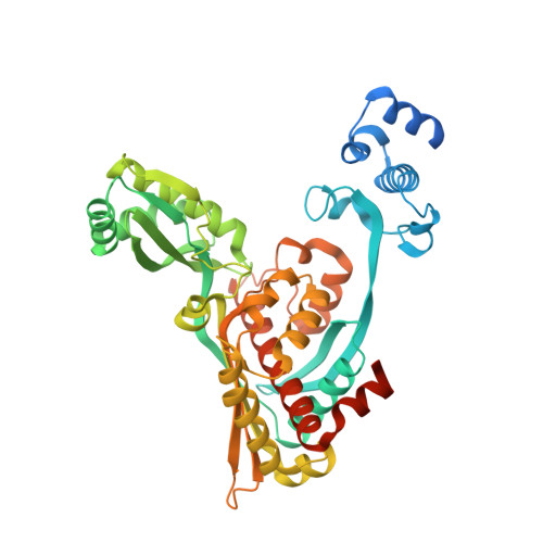
    3-hydroxy-3-methylglutaryl-CoA reductase (HMGR) catalyzes the formation of mevalonate, the committed step in the biosynthesis of sterols and isoprenoids. The activity of HMGR is controlled through synthesis, degradation and phosphorylation to maintain the concentration of mevalonate-derived products. In addition to the physiological regulation of HMGR, the human enzyme has been targeted successfully by drugs in the clinical treatment of high serum cholesterol levels. Three crystal structures of the catalytic portion of human HMGR in complexes with HMG-CoA, with HMG and CoA, and with HMG, CoA and NADP(+), provide a detailed view of the enzyme active site. Catalytic portions of human HMGR form tight tetramers. The crystal structure explains the influence of the enzyme's oligomeric state on the activity and suggests a mechanism for cholesterol sensing. The active site architecture of human HMGR is different from that of bacterial HMGR; this may explain why binding of HMGR inhibitors to bacterial HMGRs has not been reported.


  • Organizational Affiliation
    • Howard Hughes Medical Institute, Department of Biochemistry, University of Texas Southwestern Medical Center at Dallas, TX 75235-9050, USA.

Macromolecule Content 

  • Total Structure Weight: 206.78 kDa 
  • Atom Count: 12,909 
  • Modeled Residue Count: 1,614 
  • Deposited Residue Count: 1,868 
  • Unique protein chains: 1

Macromolecules

Find similar proteins by:|  3D Structure
Entity ID: 1
MoleculeChains  Sequence LengthOrganismDetailsImage
PROTEIN (HMG-COA REDUCTASE)
A, B, C, D
467Homo sapiensMutation(s): 1 
EC: 1.1.1.34
UniProt & NIH Common Fund Data Resources
Find proteins for P04035 (Homo sapiens)
Explore P04035 
Go to UniProtKB:  P04035
PHAROS:  P04035
GTEx:  ENSG00000113161 
Entity Groups
Sequence Clusters30% Identity50% Identity70% Identity90% Identity95% Identity100% Identity
UniProt GroupP04035
Sequence Annotations
Expand
Reference Sequence

Small Molecules

Ligands 3 Unique
IDChains Name / Formula / InChI Key2D Diagram3D Interactions
COA

Query on COA


Download:Ideal Coordinates CCD File
E [auth A],
H [auth B],
K [auth C],
N [auth D]
COENZYME A
C21 H36 N7 O16 P3 S
RGJOEKWQDUBAIZ-IBOSZNHHSA-N
NAP

Query on NAP


Download:Ideal Coordinates CCD File
G [auth A],
J [auth B],
L [auth C],
P [auth D]
NADP NICOTINAMIDE-ADENINE-DINUCLEOTIDE PHOSPHATE
C21 H28 N7 O17 P3
XJLXINKUBYWONI-NNYOXOHSSA-N
MAH

Query on MAH


Download:Ideal Coordinates CCD File
F [auth A],
I [auth B],
M [auth C],
O [auth D]
3-HYDROXY-3-METHYL-GLUTARIC ACID
C6 H10 O5
NPOAOTPXWNWTSH-UHFFFAOYSA-N
Binding Affinity Annotations 
IDSourceBinding Affinity
MAH BindingDB:  1DQA Ki: 23.5 (nM) from 1 assay(s)
IC50: 4000 (nM) from 1 assay(s)

Experimental Data & Validation

Experimental Data

  • Method: X-RAY DIFFRACTION
  • Resolution: 2.00 Å
  • R-Value Free:  0.197 (Depositor) 
  • R-Value Work:  0.168 (Depositor), 0.177 (DCC) 
  • R-Value Observed: 0.168 (Depositor) 
Space Group: P 1 21 1
Unit Cell:
Length ( Å )Angle ( ˚ )
a = 73.918α = 90
b = 172.63β = 117.5
c = 73.991γ = 90
Software Package:
Software NamePurpose
EPMRphasing
CNSrefinement
DENZOdata reduction
SCALEPACKdata scaling

Structure Validation

View Full Validation Report


Ligand Structure Quality Assessment 


Entry History 

Deposition Data

Revision History  (Full details and data files)

  • Version 1.0: 2000-03-08
    Type: Initial release
  • Version 1.1: 2008-04-27
    Changes: Version format compliance
  • Version 1.2: 2011-07-13
    Changes: Version format compliance
  • Version 1.3: 2018-03-14
    Changes: Database references
  • Version 1.4: 2024-02-07
    Changes: Data collection, Database references, Derived calculations