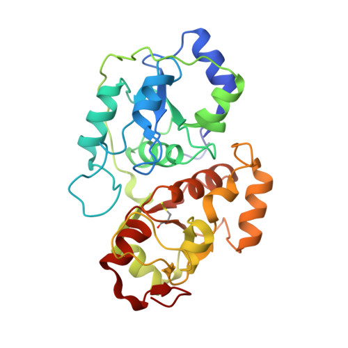
1DP2

CRYSTAL STRUCTURE OF THE COMPLEX BETWEEN RHODANESE AND LIPOATE


Experimental Data Snapshot

  • Method: X-RAY DIFFRACTION
  • Resolution: 2.01 Å
  • R-Value Free: 0.230 
  • R-Value Work: 0.170 
  • R-Value Observed: 0.170 

wwPDB Validation   3D Report Full Report


This is version 1.4 of the entry. See complete history


Literature

Specific interaction of lipoate at the active site of rhodanese.

Cianci, M.Gliubich, F.Zanotti, G.Berni, R.

(2000) Biochim Biophys Acta 1481: 103-108

  • DOI: https://doi.org/10.1016/s0167-4838(00)00114-x
  • Primary Citation of Related Structures:  
    1DP2

  • PubMed Abstract: 

    Dihydrolipoate is an acceptor of the rhodanese-bound sulfane sulfur atom, as shown by analysis of the elementary steps of the reaction catalyzed by rhodanese. The crystal structure of sulfur-substituted rhodanese complexed with the non-reactive oxidized form of lipoate has revealed that the compound is bound at the enzyme active site, with the dithiolane ring buried in the interior of the cavity and the carboxylic end pointing towards the solvent. One of the sulfur atoms of the ligand in the unproductive complex is relatively close to the sulfane sulfur bound to Cys-247, the sulfur that is transferred during the catalytic reaction. This mode of binding of lipoate is likely to mimic that of dihydrolipoate. The results presented here support the possible role of dihydrolipoate as sulfur-acceptor substrate of rhodanese in an enzymatic reaction that might serve to provide iron-sulfur proteins with inorganic sulfide.


  • Organizational Affiliation

    Department of Organic Chemistry, University of Padua, Italy.


Macromolecules
Find similar proteins by:  (by identity cutoff)  |  3D Structure
Entity ID: 1
MoleculeChains Sequence LengthOrganismDetailsImage
RHODANESE293Bos taurusMutation(s): 0 
EC: 2.8.1.1
UniProt
Find proteins for P00586 (Bos taurus)
Explore P00586 
Go to UniProtKB:  P00586
Entity Groups  
Sequence Clusters30% Identity50% Identity70% Identity90% Identity95% Identity100% Identity
UniProt GroupP00586
Sequence Annotations
Expand
  • Reference Sequence
Small Molecules
Ligands 1 Unique
IDChains Name / Formula / InChI Key2D Diagram3D Interactions
LPB
Query on LPB

Download Ideal Coordinates CCD File 
B [auth A]5-[(3S)-1,2-dithiolan-3-yl]pentanoic acid
C8 H14 O2 S2
AGBQKNBQESQNJD-ZETCQYMHSA-N
Modified Residues  1 Unique
IDChains TypeFormula2D DiagramParent
CSS
Query on CSS
A
L-PEPTIDE LINKINGC3 H7 N O2 S2CYS
Experimental Data & Validation

Experimental Data

  • Method: X-RAY DIFFRACTION
  • Resolution: 2.01 Å
  • R-Value Free: 0.230 
  • R-Value Work: 0.170 
  • R-Value Observed: 0.170 
  • Space Group: C 1 2 1
Unit Cell:
Length ( Å )Angle ( ˚ )
a = 154.36α = 90
b = 49.31β = 99.6
c = 41.68γ = 90
Software Package:
Software NamePurpose
X-PLORmodel building
X-PLORrefinement
FRAMBOdata collection
SAINTdata scaling
X-PLORphasing

Structure Validation

View Full Validation Report


Entry History 

Deposition Data

Revision History  (Full details and data files)

  • Version 1.0: 2000-12-13
    Type: Initial release
  • Version 1.1: 2008-04-27
    Changes: Version format compliance
  • Version 1.2: 2011-07-13
    Changes: Version format compliance
  • Version 1.3: 2011-10-05
    Changes: Non-polymer description
  • Version 1.4: 2017-10-04
    Changes: Refinement description