1DM0 | pdb_00001dm0

SHIGA TOXIN


Experimental Data Snapshot

  • Method: X-RAY DIFFRACTION
  • Resolution: 2.50 Å
  • R-Value Work: 
    0.206 (Depositor) 
  • R-Value Observed: 
    0.206 (Depositor) 

wwPDB Validation 3D Report Full Report

Validation slider image for 1DM0

This is version 1.6 of the entry. See complete history

Literature

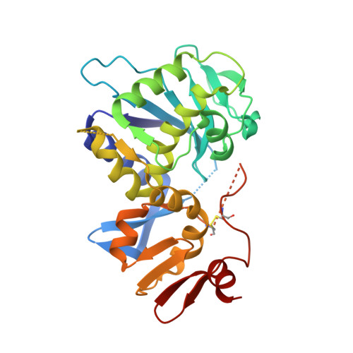
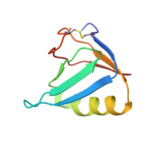
Crystal structure of the holotoxin from Shigella dysenteriae at 2.5 A resolution.

Fraser, M.E.Chernaia, M.M.Kozlov, Y.V.James, M.N.

(1994) Nat Struct Biol 1: 59-64

  • DOI: https://doi.org/10.1038/nsb0194-59
  • Primary Citation Related Structures: 
    1DM0

  • PubMed Abstract: 

    Shigella dysenteriae is the pathogen responsible for the severe form of dysentery in humans. It produces Shiga toxin, the prototype of a family of closely related bacterial protein toxins. We have determined the structure of the holotoxin, an AB5 hexamer, by X-ray crystallography. The five B subunits form a pentameric ring, encircling a helix at the carboxy terminus of the A subunit. The A subunit interacts with the B pentamer via this C-terminal helix and a four-stranded mixed beta-sheet. The fold of the rest of the A subunit is similar to that of the A chain of the plant toxin ricin; both are N-glycosidases. However, the active site in the bacterial holotoxin is blocked by a segment of polypeptide chain. These residues of the A subunit would be released as part of the activation mechanism of the toxin.


  • Organizational Affiliation
    • Department of Biochemistry, University of Alberta, Edmonton, Canada.

Macromolecule Content 

  • Total Structure Weight: 140.11 kDa 
  • Atom Count: 9,538 
  • Modeled Residue Count: 1,216 
  • Deposited Residue Count: 1,264 
  • Unique protein chains: 2

Macromolecules

Find similar proteins by:|  3D Structure
Entity ID: 1
MoleculeChains  Sequence LengthOrganismDetailsImage
SHIGA TOXIN A SUBUNITA,
B [auth L]
287Shigella dysenteriaeMutation(s): 0 
EC: 3.2.2.22
UniProt
Find proteins for Q9FBI2 (Shigella dysenteriae)
Explore Q9FBI2 
Go to UniProtKB:  Q9FBI2
Entity Groups
Sequence Clusters30% Identity50% Identity70% Identity90% Identity95% Identity100% Identity
UniProt GroupQ9FBI2
Sequence Annotations
Expand
Reference Sequence
Find similar proteins by:|  3D Structure
Entity ID: 2
MoleculeChains  Sequence LengthOrganismDetailsImage
SHIGA TOXIN B SUBUNIT69Shigella dysenteriaeMutation(s): 0 
UniProt
Find proteins for Q7BQ98 (Shigella dysenteriae)
Explore Q7BQ98 
Go to UniProtKB:  Q7BQ98
Entity Groups
Sequence Clusters30% Identity50% Identity70% Identity90% Identity95% Identity100% Identity
UniProt GroupQ7BQ98
Sequence Annotations
Expand
Reference Sequence

Experimental Data & Validation

Experimental Data

  • Method: X-RAY DIFFRACTION
  • Resolution: 2.50 Å
  • R-Value Work:  0.206 (Depositor) 
  • R-Value Observed: 0.206 (Depositor) 
Space Group: P 21 21 21
Unit Cell:
Length ( Å )Angle ( ˚ )
a = 133.05α = 90
b = 147.46β = 90
c = 83.04γ = 90
Software Package:
Software NamePurpose
TNTrefinement
BIOMOLdata reduction
BRUTEmodel building
MLPHAREphasing
DEMONmodel building
X-PLORrefinement
WEISdata scaling
WEISdata reduction
BIOMOLdata scaling
BRUTEphasing
DEMONphasing

Structure Validation

View Full Validation Report


Entry History 

Deposition Data

Revision History  (Full details and data files)

  • Version 1.0: 1999-12-30
    Type: Initial release
  • Version 1.1: 2008-04-27
    Changes: Version format compliance
  • Version 1.2: 2011-07-13
    Changes: Version format compliance
  • Version 1.3: 2017-10-04
    Changes: Refinement description
  • Version 1.4: 2019-07-24
    Changes: Data collection, Refinement description
  • Version 1.5: 2019-08-14
    Changes: Data collection
  • Version 1.6: 2024-10-16
    Changes: Data collection, Database references, Structure summary