1DKS | pdb_00001dks

CKSHS1: HUMAN CYCLIN DEPENDENT KINASE SUBUNIT, TYPE 1 IN COMPLEX WITH PHOSPHATE


Experimental Data Snapshot

  • Method: X-RAY DIFFRACTION
  • Resolution: 3.20 Å
  • R-Value Free: 
    0.290 (Depositor) 
  • R-Value Work: 
    0.183 (Depositor) 
  • R-Value Observed: 
    0.183 (Depositor) 

wwPDB Validation 3D Report Full Report

Validation slider image for 1DKS

This is version 1.4 of the entry. See complete history

Literature

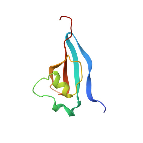
Crystal structure of the human cell cycle protein CksHs1: single domain fold with similarity to kinase N-lobe domain.

Arvai, A.S.Bourne, Y.Hickey, M.J.Tainer, J.A.

(1995) J Mol Biology 249: 835-842

  • DOI: https://doi.org/10.1006/jmbi.1995.0341
  • Primary Citation Related Structures: 
    1DKS, 1DKT

  • PubMed Abstract: 

    The structure of the human CksHs1 homolog of the yeast cell-cycle regulatory proteins suc1 and CKS1, which bind to the catalytic subunit of the cyclin-dependent kinases (Cdks) and are essential for yeast cell-cycle progression in vivo, has been determined at 2.9 A resolution. The CksHs1 single polypeptide domain fold, which consists of a four-stranded beta-sheet flanked by two alpha-helices, is dramatically different from the subunit conformation and assembly of the homologous CksHs2, but strikingly similar to the Cdk N-lobe domain fold. The CksHs1 structure identifies sequence-conserved residues Glu61 to His65 as a novel beta-hinge region that folds back to form a beta-hairpin with CksHs1 subunit, whereas this hinge is unfolded to form an extended beta-strand exchange between two CksHs2 subunits. Phosphate and the phosphate analog metavanadate bind CksHs1 in a shallow pocket and interact with five conserved residues (Lys11, Arg20, Ser51, Trp54 and Arg71) suggesting a specific Cks recognition site for a phosphorylated Cdk residue. The dramatic changes to the Cks fold, assembly and exposed conserved surface brought about by switching between the bent and extended hinge conformations are potentially important for the functions of this Cks homolog and could explain conflicting activities inferred from different types of genetic experiments.


  • Organizational Affiliation
    • Department of Molecular Biology, Scripps Research Institute, La Jolla, CA 92037, USA.

Macromolecule Content 

  • Total Structure Weight: 19.55 kDa 
  • Atom Count: 1,331 
  • Modeled Residue Count: 149 
  • Deposited Residue Count: 158 
  • Unique protein chains: 1

Macromolecules

Find similar proteins by:|  3D Structure
Entity ID: 1
MoleculeChains  Sequence LengthOrganismDetailsImage
CYCLIN DEPENDENT KINASE SUBUNIT, TYPE 1
A, B
79Homo sapiensMutation(s): 0 
UniProt & NIH Common Fund Data Resources
Find proteins for P61024 (Homo sapiens)
Explore P61024 
Go to UniProtKB:  P61024
PHAROS:  P61024
GTEx:  ENSG00000173207 
Entity Groups
Sequence Clusters30% Identity50% Identity70% Identity90% Identity95% Identity100% Identity
UniProt GroupP61024
Sequence Annotations
Expand
Reference Sequence

Small Molecules

Experimental Data & Validation

Experimental Data

  • Method: X-RAY DIFFRACTION
  • Resolution: 3.20 Å
  • R-Value Free:  0.290 (Depositor) 
  • R-Value Work:  0.183 (Depositor) 
  • R-Value Observed: 0.183 (Depositor) 
Space Group: P 61 2 2
Unit Cell:
Length ( Å )Angle ( ˚ )
a = 94α = 90
b = 94β = 90
c = 137.4γ = 120
Software Package:
Software NamePurpose
X-PLORmodel building
X-PLORrefinement
XDSdata reduction
X-PLORphasing

Structure Validation

View Full Validation Report


Entry History 

Deposition Data

Revision History  (Full details and data files)

  • Version 1.0: 1996-03-08
    Type: Initial release
  • Version 1.1: 2008-03-03
    Changes: Version format compliance
  • Version 1.2: 2011-07-13
    Changes: Version format compliance
  • Version 1.3: 2017-11-29
    Changes: Derived calculations, Other
  • Version 1.4: 2024-02-07
    Changes: Data collection, Database references, Derived calculations