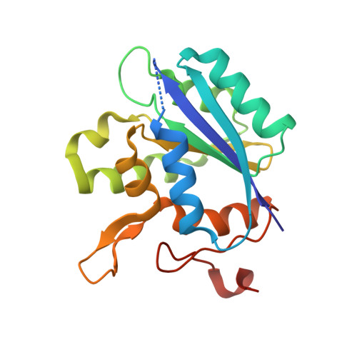
1DI6

1.45 A CRYSTAL STRUCTURE OF THE MOLYBDENUMM COFACTOR BIOSYNTHESIS PROTEIN MOGA FROM ESCHERICHIA COLI


Experimental Data Snapshot

  • Method: X-RAY DIFFRACTION
  • Resolution: 1.45 Å
  • R-Value Free: 0.213 
  • R-Value Work: 0.187 

wwPDB Validation   3D Report Full Report


This is version 1.4 of the entry. See complete history


Literature

Crystal structure of the gephyrin-related molybdenum cofactor biosynthesis protein MogA from Escherichia coli.

Liu, M.T.Wuebbens, M.M.Rajagopalan, K.V.Schindelin, H.

(2000) J Biol Chem 275: 1814-1822

  • DOI: https://doi.org/10.1074/jbc.275.3.1814
  • Primary Citation of Related Structures:  
    1DI6, 1DI7

  • PubMed Abstract: 

    Molybdenum cofactor (Moco) biosynthesis is an evolutionarily conserved pathway in archaea, eubacteria, and eukaryotes, including humans. Genetic deficiencies of enzymes involved in this biosynthetic pathway trigger an autosomal recessive disease with severe neurological symptoms, which usually leads to death in early childhood. The MogA protein exhibits affinity for molybdopterin, the organic component of Moco, and has been proposed to act as a molybdochelatase incorporating molybdenum into Moco. MogA is related to the protein gephyrin, which, in addition to its role in Moco biosynthesis, is also responsible for anchoring glycinergic receptors to the cytoskeleton at inhibitory synapses. The high resolution crystal structure of the Escherichia coli MogA protein has been determined, and it reveals a trimeric arrangement in which each monomer contains a central, mostly parallel beta-sheet surrounded by alpha-helices on either side. Based on structural and biochemical data, a putative active site was identified, including two residues that are essential for the catalytic mechanism.


  • Organizational Affiliation

    Department of Biochemistry and Cell Biology, State University of New York, Stony Brook, New York 11794-5215, USA.


Macromolecules
Find similar proteins by:  (by identity cutoff)  |  3D Structure
Entity ID: 1
MoleculeChains Sequence LengthOrganismDetailsImage
MOLYBDENUM COFACTOR BIOSYNTHETIC ENZYME195Escherichia coliMutation(s): 1 
EC: 2.7.7.75
UniProt
Find proteins for P0AF03 (Escherichia coli (strain K12))
Explore P0AF03 
Go to UniProtKB:  P0AF03
Entity Groups  
Sequence Clusters30% Identity50% Identity70% Identity90% Identity95% Identity100% Identity
UniProt GroupP0AF03
Sequence Annotations
Expand
  • Reference Sequence
Experimental Data & Validation

Experimental Data

  • Method: X-RAY DIFFRACTION
  • Resolution: 1.45 Å
  • R-Value Free: 0.213 
  • R-Value Work: 0.187 
  • Space Group: P 63
Unit Cell:
Length ( Å )Angle ( ˚ )
a = 65.3α = 90
b = 65.3β = 90
c = 65.3γ = 120
Software Package:
Software NamePurpose
SHARPphasing
REFMACrefinement
X-PLORrefinement
DENZOdata reduction
SCALEPACKdata scaling

Structure Validation

View Full Validation Report


Entry History 

Deposition Data

Revision History  (Full details and data files)

  • Version 1.0: 2000-01-19
    Type: Initial release
  • Version 1.1: 2008-04-27
    Changes: Version format compliance
  • Version 1.2: 2011-07-13
    Changes: Derived calculations, Version format compliance
  • Version 1.3: 2021-11-03
    Changes: Database references, Derived calculations
  • Version 1.4: 2024-02-07
    Changes: Data collection