Long-range structural effects in a second-site revertant of a mutant dihydrofolate reductase.
Brown, K.A., Howell, E.E., Kraut, J.(1993) Proc Natl Acad Sci U S A 90: 11753-11756
- PubMed: 8265622 Search on PubMedSearch on PubMed Central
- DOI: https://doi.org/10.1073/pnas.90.24.11753
- Primary Citation Related Structures:
1DHI, 1DHJ - PubMed Abstract:
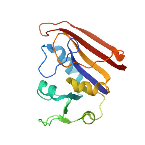
X-ray crystal structures have been determined for a second-site revertant (Asp-27-->Ser, Phe-137-->Ser; D27S/F137S) and both component single-site mutants of Escherichia coli dihydrofolate reductase. The primary D27S mutation, located in the substrate binding pocket, greatly reduces catalytic activity as compared to the wild-type enzyme. The additional F137S mutation, which partially restores catalytic activity, is located on the surface of the molecule, well outside of the catalytic center and approximately 15 A from residue 27. Comparison of kinetic data for the single-site F137S mutant, specifically constructed as a control, and for the double-mutant enzymes indicates that the effects of the F137S and D27S mutations on catalysis are nonadditive. This result suggests that the second-site mutation might mediate its effects through a structural perturbation propagated along the polypeptide backbone. To investigate the mechanism by which the F137S substitution elevates the catalytic activity of D27S we have determined the structure of the D27S/F137S double mutant. We also present a rerefined structure for the original D27S mutant and a preliminary structural interpretation for the F137S single-site mutant. We find that while either single mutant shows little more than a simple side-chain substitution, the double mutant undergoes an extended structural perturbation, which is propagated between these two widely separated sites via the helix alpha B.
- Department of Chemistry, University of California at San Diego, La Jolla 92093.
Organizational Affiliation: