1DGL

LECTIN FROM DIOCLEA GRANDIFLORA COMPLEXED TO TRIMANNOSIDE


Experimental Data Snapshot

  • Method: X-RAY DIFFRACTION
  • Resolution: 2.40 Å
  • R-Value Free: 0.252 
  • R-Value Work: 0.189 
  • R-Value Observed: 0.189 

wwPDB Validation   3D Report Full Report


This is version 2.0 of the entry. See complete history


Literature

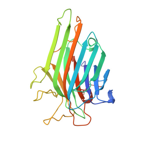
Crystal structure of the lectin from Dioclea grandiflora complexed with core trimannoside of asparagine-linked carbohydrates.

Rozwarski, D.A.Swami, B.M.Brewer, C.F.Sacchettini, J.C.

(1998) J Biol Chem 273: 32818-32825

  • DOI: https://doi.org/10.1074/jbc.273.49.32818
  • Primary Citation of Related Structures:  
    1DGL

  • PubMed Abstract: 
  • The seed lectin from Dioclea grandiflora (DGL) has recently been shown to possess high affinity for 3, 6-di-O-(alpha-D-mannopyranosyl)-alpha-D-mannopyranose, the core trimannoside of asparagine-linked carbohydrates, but lower affinity for biantennary complex carbohydrates ...

    The seed lectin from Dioclea grandiflora (DGL) has recently been shown to possess high affinity for 3, 6-di-O-(alpha-D-mannopyranosyl)-alpha-D-mannopyranose, the core trimannoside of asparagine-linked carbohydrates, but lower affinity for biantennary complex carbohydrates. In the previous paper, the thermodynamics of DGL binding to deoxy analogs of the core trimannoside and to a biantennary complex carbohydrate were determined by isothermal titration microcalorimetry. The data suggest that DGL recognizes specific hydroxyl groups of the trimannoside similar to that of the jack bean lectin concanavalin A (ConA) (Gupta, D. Dam, T. K., Oscarson, S., and Brewer, C. F. (1997) J. Biol. Chem. 272, 6388-6392). However, the thermodynamics of DGL binding to certain deoxy analogs and to the complex carbohydrate are different from that of ConA. In the present paper, the x-ray crystal structure of DGL complexed to the core trimannoside was determined to a resolution of 2.6 A. The overall structure of the DGL complex is similar to the structure of the ConA-trimannoside complex (Naismith, J. H., and Field, R. A. (1996) J. Biol. Chem. 271, 972-976). The location and conformation of the bound trimannoside as well as its hydrogen-bonding interactions in both complexes are nearly identical. However, differences exist in the location of two loops outside of the respective binding sites containing residues 114-125 and 222-227. The latter residues affect the location of a network of hydrogen-bonded water molecules that interact with the trisaccharide. Differences in the arrangement of ordered water molecules in the binding site and/or protein conformational differences outside of the binding site may account for the differences in the thermodynamics of binding of the two lectins to deoxy analogs of the trimannoside. Molecular modeling studies suggest how DGL discriminates against binding the biantennary complex carbohydrate relative to ConA.


    Related Citations: 
    • Structural Basis of Trimannoside Recognition by Concanavalin A
      Naismith, J.H., Field, R.A.
      (1996) J Biol Chem 271: 972

    Organizational Affiliation

    Department of Biochemistry, Albert Einstein College of Medicine, Bronx, New York 10461, USA.


Macromolecules
Find similar proteins by:  (by identity cutoff)  |  3D Structure
Entity ID: 1
MoleculeChainsSequence LengthOrganismDetailsImage
LECTINA, B237Macropsychanthus grandiflorusMutation(s): 0 
UniProt
Find proteins for P08902 (Dioclea grandiflora)
Explore P08902 
Go to UniProtKB:  P08902
Entity Groups  
Sequence Clusters30% Identity50% Identity70% Identity90% Identity95% Identity100% Identity
UniProt GroupP08902
Protein Feature View
Expand
  • Reference Sequence
Oligosaccharides

Help

Entity ID: 2
MoleculeChainsChain Length2D DiagramGlycosylation3D Interactions
alpha-D-mannopyranose-(1-3)-[alpha-D-mannopyranose-(1-6)]methyl alpha-D-mannopyranosideC, D 3N/A Oligosaccharides Interaction
Glycosylation Resources
GlyTouCan:  G27351BJ
GlyCosmos:  G27351BJ
Small Molecules
Ligands 2 Unique
IDChainsName / Formula / InChI Key2D Diagram3D Interactions
MN
Query on MN

Download Ideal Coordinates CCD File 
F [auth A],
H [auth B]
MANGANESE (II) ION
Mn
WAEMQWOKJMHJLA-UHFFFAOYSA-N
 Ligand Interaction
CA
Query on CA

Download Ideal Coordinates CCD File 
E [auth A],
G [auth B]
CALCIUM ION
Ca
BHPQYMZQTOCNFJ-UHFFFAOYSA-N
 Ligand Interaction
Experimental Data & Validation

Experimental Data

  • Method: X-RAY DIFFRACTION
  • Resolution: 2.40 Å
  • R-Value Free: 0.252 
  • R-Value Work: 0.189 
  • R-Value Observed: 0.189 
  • Space Group: P 31 2 1
Unit Cell:
Length ( Å )Angle ( ˚ )
a = 92.11α = 90
b = 92.11β = 90
c = 202.82γ = 120
Software Package:
Software NamePurpose
X-PLORmodel building
X-PLORrefinement
SADIEdata reduction
SAINTdata reduction
SADIEdata scaling
SAINTdata scaling
X-PLORphasing

Structure Validation

View Full Validation Report


Entry History 

Deposition Data

Revision History  (Full details and data files)

  • Version 1.0: 1999-06-01
    Type: Initial release
  • Version 1.1: 2008-03-24
    Changes: Version format compliance
  • Version 1.2: 2011-07-13
    Changes: Derived calculations, Version format compliance
  • Version 2.0: 2020-07-29
    Type: Remediation
    Reason: Carbohydrate remediation
    Changes: Atomic model, Data collection, Database references, Derived calculations, Other, Structure summary