1DDS

MOLECULE: DIHYDROFOLATE REDUCTASE (E.C.1.5.1.3) COMPLEXED WITH METHOTREXATE


Experimental Data Snapshot

  • Method: X-RAY DIFFRACTION
  • Resolution: 2.20 Å
  • R-Value Free: 0.200 
  • R-Value Work: 0.177 
  • R-Value Observed: 0.177 

wwPDB Validation   3D Report Full Report


Ligand Structure Quality Assessment 


This is version 1.3 of the entry. See complete history


Literature

The effect of denaturants on protein structure.

Dunbar, J.Yennawar, H.P.Banerjee, S.Luo, J.Farber, G.K.

(1997) Protein Sci 6: 1727-1733

  • DOI: https://doi.org/10.1002/pro.5560060813
  • Primary Citation of Related Structures:  
    1DDR, 1DDS, 1RBW, 1RBX

  • PubMed Abstract: 

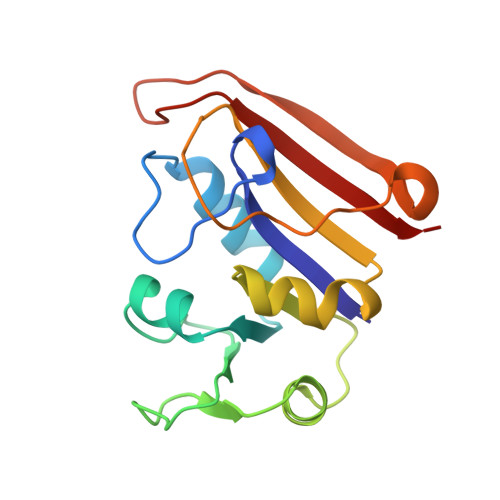
    Virtually all studies of the protein-folding reaction add either heat, acid, or a chemical denaturant to an aqueous protein solution in order to perturb the protein structure. When chemical denaturants are used, very high concentrations are usually necessary to observe any change in protein structure. In a solution with such high denaturant concentrations, both the structure of the protein and the structure of the solvent around the protein can be altered. X-ray crystallography is the obvious experimental technique to probe both types of changes. In this paper, we report the crystal structures of dihydrofolate reductase with urea and of ribonuclease A with guanidinium chloride. These two classic denaturants have similar effects on the native structure of the protein. The most important change that occurs is a reduction in the overall thermal factor. These structures offer a molecular explanation for the reduction in mobility. Although the reduction is observed only with the native enzyme in the crystal, a similar decrease in mobility has also been observed in the unfolded state in solution (Makhatadze G, Privalov PL. 1992. Protein interactions with urea and guanidinium chloride: A calorimetric study.


  • Organizational Affiliation

    Department of Biochemistry and Molecular Biology, Pennsylvania State University, University Park 16802, USA.


Macromolecules
Find similar proteins by:  (by identity cutoff)  |  3D Structure
Entity ID: 1
MoleculeChains Sequence LengthOrganismDetailsImage
DIHYDROFOLATE REDUCTASE
A, B
159Escherichia coliMutation(s): 0 
EC: 1.5.1.3
UniProt
Find proteins for P0ABQ4 (Escherichia coli (strain K12))
Explore P0ABQ4 
Go to UniProtKB:  P0ABQ4
Entity Groups  
Sequence Clusters30% Identity50% Identity70% Identity90% Identity95% Identity100% Identity
UniProt GroupP0ABQ4
Sequence Annotations
Expand
  • Reference Sequence
Experimental Data & Validation

Experimental Data

  • Method: X-RAY DIFFRACTION
  • Resolution: 2.20 Å
  • R-Value Free: 0.200 
  • R-Value Work: 0.177 
  • R-Value Observed: 0.177 
  • Space Group: P 61
Unit Cell:
Length ( Å )Angle ( ˚ )
a = 93.181α = 90
b = 93.185β = 90
c = 73.886γ = 120
Software Package:
Software NamePurpose
X-PLORmodel building
X-PLORrefinement
TEXSANdata reduction
X-PLORphasing

Structure Validation

View Full Validation Report


Ligand Structure Quality Assessment 


Entry History 

Deposition Data

Revision History  (Full details and data files)

  • Version 1.0: 1995-10-15
    Type: Initial release
  • Version 1.1: 2008-03-24
    Changes: Version format compliance
  • Version 1.2: 2011-07-13
    Changes: Version format compliance
  • Version 1.3: 2024-02-07
    Changes: Data collection, Database references, Derived calculations, Other, Structure summary