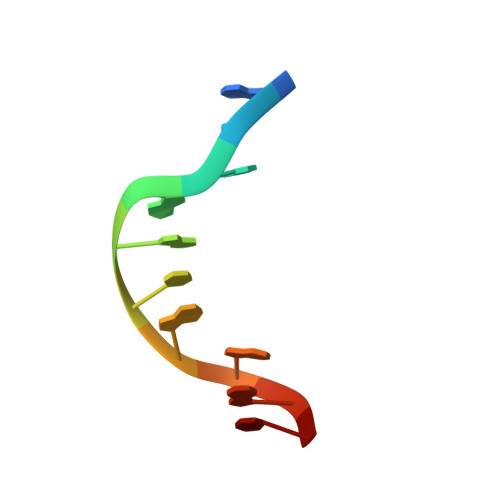
Solution structure of [d(ATGAGCGAATA)]2. Adjacent G:A mismatches stabilized by cross-strand base-stacking and BII phosphate groups.
Chou, S.H., Cheng, J.W., Reid, B.R.(1992) J Mol Biol 228: 138-155
- PubMed: 1447778 Search on PubMed
- DOI: https://doi.org/10.1016/0022-2836(92)90497-8
- Primary Citation Related Structures:
1D69 - PubMed Abstract:
The solution structure of a rather unusual B-form duplex [d(ATGAGCGAATA)]2 has been determined using two-dimensional nuclear magnetic resonance (2D-NMR) and distance geometry methods. This sequence forms a stable ten base-pair B-form duplex with 3' overhangs and two pairs of adjacent G:A mismatches paired via a sheared hydrogen-bonding scheme. All non-exchangeable protons, including the stereo-specific H-5'S/H-5'R of the 3G and 7G residues, were assigned by 2D-NMR. The phosphorus spectrum was assigned using heteronuclear correlation with H-3' and H-4' reasonances. The complete assignments reveal several unusual nuclear Overhauser enhancements (NOEs) and unusual chemical shifts for the neighboring G:A mismatch pairs and their adjacent nucleotides. Inter-proton distances were derived from time-dependent NOEs and used to generate initial structures, which were further refined by iterative back-calculation of the two-dimensional nuclear Overhauser enhancement spectra; 22 final structures were calculated from the refined distance bounds. All these final structures exhibit fully wound helical structures with small penalty values against the refined distance bounds and small pair-wise root-mean-square deviation values (typically 0.5 A to 0.9 A). The two helical strands exchange base stacking at both of the two G:A mismatch sites, resulting in base stacking down each side rather than down each strand of the twisted duplex. Very large twist angles (77 degrees) were found at the G:A mismatch steps. All the final structures were found to have BII phosphate conformations at the adjacent G:A mismatch sites, consistent with observed downfield 31P chemical shifts and Monte-Carlo conformational search results. Our results support the hypothesis that 31P chemical shifts are related to backbone torsion angles. These BII phosphate conformations in the adjacent G:A mismatch step suggest that hydrogen bonding of the G:A pair G-NH2 to a nearby phosphate oxygen atom is unlikely. The unusual structure of the duplex may be stabilized by strong interstrand base stacking as well as intrastrand stacking, as indicated by excellent base overlap within the mismatch stacks.
- Howard Hughes Medical Institute, University of Washington, Seattle 98195.
Organizational Affiliation: